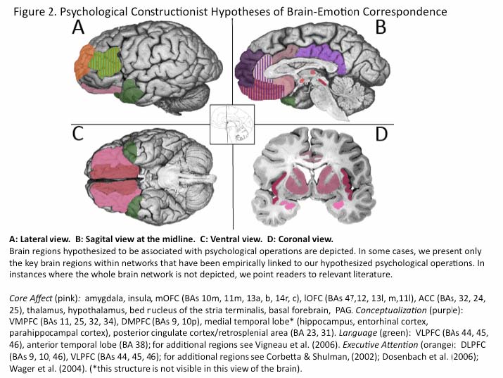
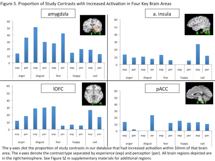
>

The brain basis of emotion: A meta-analytic review

Kristen A. Lindquist, Tor D. Wager, Hedy Kober, Eliza Bliss-Moreau, and Lisa Feldman Barrett

Address correspondence to: Kristen A. Lindquist

Contributing Authors

Kristen A. Lindquist, Ph.D. Postdoctoral Fellow Mind/Brain/Behavior Initiative, Harvard University Department of Neurology Massachusetts General Hospital, East CNY 149, Suite 10.022 Charlestown, MA 02129

lindqukr@nmr.mgh.harvard.edu http://www.nmr.mgh.harvard.edu/~lindqukr/

Tor. D. Wager, Ph.D. Associate Professor Department of Psychology and Neuroscience University of Colorado, Boulder

tor.wager@colorado.edu http://www.psych.colorado.edu/~tor/

Hedy Kober, Ph.D. Assistant Professor Department of Psychiatry Yale University School of Medicine 34 Park St. Rm. S111 New Haven, CT 06519

hedy.kober@yale.edu http://www.columbia.edu/~hk384/

Eliza Bliss-Moreau, Ph.D. Postdoctoral Fellow Department of Psychiatry and Behavioral Sciences California National Primate Research Center University of California, Davis One Shields Avenue Davis, CA 95616

eblissmoreau@ucdavis.edu

Lisa Feldman Barrett, Ph.D. Distinguished Professor Department of Psychology Northeastern University Boston, MA 02115

l.barrett@neu.edu http://www.affective-science.org/

Abstract: Researchers have wondered how the brain creates emotions since the early days of psychological science. With a surge of studies in affective neuroscience in recent decades, scientists are poised to answer this question. In this paper, we present a meta-analytic summary of the human neuroimaging literature on emotion. We compare the locationist approach (i.e., the hypothesis that discrete emotion categories consistently and specifically correspond to distinct brain regions) with the psychological constructionist approach (i.e., the hypothesis that discrete emotion categories are constructed of more general brain networks not specific to those categories) to better understand the brain basis of emotion. We review both locationist and psychological constructionist hypotheses of brain-emotion correspondence and report metaanalytic findings bearing on these hypotheses. Overall, we found little evidence that discrete emotion categories can be consistently and specifically localized to distinct brain regions. Instead, we found evidence that is consistent with a psychological constructionist approach to the mind: a set of interacting brain regions commonly involved in basic psychological operations of both an emotional and non-emotional nature are active during emotion experience and perception across a range of discrete emotion categories.

Keywords: emotion, psychological construction, discrete emotion, emotion experience, emotion perception, meta-analysis, neuroimaging

1. Introduction

William James framed the question of emotion-brain correspondence when he wrote, “…of two things concerning the emotions, one must be true. Either separate and special centres, affected to them alone, are their brain-seat, or else they correspond to processes occurring in the motor and sensory centres already assigned…” (1890, p. 473). In this paper, we statistically summarize the last fifteen years of neuroimaging research on emotion in an attempt to determine which of these alternatives is correct. We examine the utility of two different models of emotion that have each existed since the beginning of psychology.

2. A Locationist Account of the Brain Basis of Emotion

A locationist account of emotion assumes that the category emotion and individual categories such as anger, disgust, fear, happiness, sadness (and perhaps a few others) are respected by the body and brain (see Barrett, 2006a for a discussion). The guiding hypothesis of this natural kind model (Barrett, 2006a) or modal model (Barrett, Ochsner, & Gross, 2007) of emotion is that different emotion categories refer to states with endowed motivational characteristics that drive cognition and behavior. It is assumed that these states are biologically basic, inherited, and cannot be broken down into more basic psychological components (Izard, in press; Ekman & Cordaro, in press; Panksepp & Watt, in press). Despite these common assumptions, there is variability in how different researchers define emotions as natural kinds. Some theorists emphasize the universal characteristics of emotion categories, suggesting that each emotion category (e.g., anger) refers to a “family” of states that share a distinctive universal signal (e.g., facial behavior), physiology, antecedent events, subjective experience, and accompanying thoughts and memories (e.g., Ekman & Cordano, in press). In this view, emotions can be shaped by culture and learning, but all humans possess the capacity to experience and perceive the same core set of emotion categories. Other theorists take a developmental approach and argue that all infants are born with a set of “first order emotions” that are evolutionarily given reactions (including feelings, motivations and behaviors) to specific stimuli (e.g., Izard, in press). First order emotions form the core of the more elaborate “emotion schemas” that develop with age and learning and consist of complex combinations of emotions, cognitions, and behaviors. Still other theorists emphasize the evolutionary aspect of emotion categories, and argue that emotions are specific behavioral adaptations that are shared with other mammalian species and passed down through phylogeny (e.g., Panksepp, 1998; 2007; Panksepp & Watt, in press). Some models taking an “appraisal” approach to emotion also draw on natural kind assumptions about emotions (cf. Barrett, 2006a) by hypothesizing that dedicated cognitive mechanisms automatically make meaning of a stimulus and trigger the corresponding discrete emotion (e.g., Roseman, 1984; Scherer et al. 2003). Relatively little work from an appraisal perspective has investigated the brain basis of emotion (although see Sander et al 2003; 2007), so we do not discuss appraisal models further in this paper.

All natural kind models share the assumption that different emotion categories have their roots in distinct mechanisms in the brain and body. The mechanisms underlying discrete emotion categories have been discussed as particular gross anatomical locations (e.g., Calder, 2003, Ekman, 1999) or networks (e.g., Izard, in press; Panksepp, 1998) in the brain. These models constitute a locationist account of emotion because they hypothesize that all mental states belonging to the same emotion category (e.g., fear) are produced by activity that is consistently and specifically associated with an architecturally defined brain locale (see sections 5.1-5.4 below)1 or anatomically defined networks of locales that are inherited and shared with other mammalian species (Panksepp, 1998; Panksepp & Watt, in press). Not all natural kind models are locationist, however; for example, some models propose that each discrete emotion is triggered by an inherited mechanism (e.g., an “affect program;” Tomkins, 1962; Ekman & Cordaro, in press) that does not necessarily correspond to a particular brain locale but rather to a specific pattern of autonomic nervous system activity. Much of the contemporary research on emotion makes locationist assumptions and in this paper we focus on the models that hypothesize single brain regions to be consistently and specifically associated with different emotion categories because they represent the most frequent hypothesis that has been tested in the cognitive neuroscience literature. We discuss specific predictions of the locationist approach in section 5, Testing Hypotheses of Brain-Emotion Correspondence (also see Figure 1).

3. A Psychological Constructionist Account of the Brain Basis of Emotion

A psychological constructionist account of emotion assumes that emotions are psychological events that emerge out of more basic psychological operations that are not specific to emotion. In this view, mental categories like anger, sadness, fear, etc. are not respected by the brain (nor are emotion, perception or cognition, for that matter; Barrett, 2009; Duncan & Barrett, 2007; Pessoa, 2008). A psychological constructionist approach to emotion is as old as the locationist approach, at least in its nascent form (e.g., Wundt, James and other early psychologists were psychological constructionists, see Gendron & Barrett, 2009). Our contemporary psychological constructionist approach shares much in common with cognitive neuroscience approaches arguing that basic psychological operations are common across diverse task domains (Cole & Schneider, 2007; Dosenbach, et al. 2006; Smith, et al. 2009; Wager et al. 2005; Wager & Smith, 2003; van Snellenberg & Wager, 2009). Like the neural context hypothesis, it assumes that the psychological function of individual brain regions is determined, in part, by the network of brain regions it is firing with (McIntosh, 2004). It is also consistent with recent evidence that largescale networks intrinsic to the brain interact to produce psychological events (Seeley et al 2007; Smith et al 2009; Spreng et al. 2010; see Deco, Jirsa & MacIntosh, 2011 for a review). In philosophy of mind, it is consistent with both a token identity and supervenience approach to mind-brain correspondence (Barrett, in press) and the mental mechanisms approach (Bechtel, 2007). We discuss the psychological constructionist view in a bit more detail because it is unfamiliar to many readers.

In our psychological constructionist model, called the “conceptual act model,” emotions emerge when people make meaning out of sensory input from the body and from the world using knowledge of prior experiences. Emotions are “situated conceptualizations” (cf. Barsalou, 2003) because the emerging meaning is tailored to the immediate environment and prepares the person to respond to sensory input in a way that is tailored to the situation (Barrett, 2006b). “Conceptual acts” could also be called “perceptual acts” because they are thought to emerge in consciousness just as visual and auditory percepts do when sensory input is automatically and effortlessly made meaningful using knowledge from prior experience. The idea is that the brain makes an initial prediction about the meaning of the sensory array in context (Bar, 2007), and the error between this initial top-down prediction and the sensory activity is quickly minimized (Friston, 2010) to produce a unified conscious field.

In psychological construction, all mental states, whether they are experienced as an instance of a discrete emotion category or not, are realized by more basic psychological operations or “ingredients” of the mind. The goal of psychology is to identify these psychological operations as “psychological primitives,” or the most basic psychological descriptions that cannot be further reduced to anything else mental (because at that point they would describe biological mechanisms). These basic psychological operations are not functionally specific to any discrete emotion category, or even to the category emotion itself. Instead, they are functionally selective for emotion on certain occasions. Because our own model is relatively new, our current model has not identified the most primitive level of psychological description, and it is not yet possible to definitively claim what the most basic psychological operations of the mind are. What we propose is a set of basic domains of psychological function that are a first approximation in the trajectory of a longer research program to identify psychological primitives; these will no doubt be refined as research proceeds in the coming years.

One operation in all psychological constructionist models of emotion is some form of sensory input from the body, such as raw somatic, visceral, vascular and motor cues (James, 1884), arousal (Duffy, 1957; Mandler, 1975; 1990; Schachter & Singer, 1962), or affect (Wundt, 1897; Harlow & Stagner, 2932; Hunt, 1941; now called core affect; Barrett 2006b; Barrett & Bliss-Moreau, 2009; Russell, 2003; Russell & Barrett, 1999). In our psychological constructionist view, we refer to this basic psychological domain as “core affect.” In psychology, the word “affect” is used to mean anything that is emotional. Core affect, on the other hand, is a term used to describe the mental representation of bodily changes that are sometimes experienced as feelings of hedonic pleasure and displeasure with some degree of arousal (Barrett & Bliss-Moreau, 2009; Russell & Barrett, 1999; Russell, 2003). Core affect is realized, in part, by visceral control systems that help organisms deal with motivationally salient stimuli in the environment. A functioning peripheral nervous system is not necessary for a person to experience a core affective state (e.g., Critchley et al. 2001) as long as they have some prior experiences to provide them with central nervous system representations of bodily states. However, in healthy individuals, core affect is usually accompanied by somatovisceral, kinesthetic, proprioceptive, and neurochemical fluctuations that take place within the core of body and are represented in the brain. Changes in core affect are a homeostatic barometer – the body’s way of representing whether objects in the environment are valuable or not in a given context. The concept of core affect shares much in common with the idea that bodily cues constitute a core ingredient in mental life (e.g., the idea that being embodied is essential to consciousness; Craig, 2009; Damasio, 1999; the idea that feelings are a common currency for valuation of objects in the world; Cabanac, 2002). We assume that core affect is not psychologically meaningful unless it is attached to an object; it is made meaningful via a second basic psychological operation, described below.

Definitions

Natural kind approach. A theoretical framework for understanding the ontology of emotions. The natural kinds approach assumes that emotion categories like anger, sadness, fear, etc. map on to biological categories that are given by the brain and body and cannot be reduced to more basic psychological parts.

Locationist approach. Many national kind models of emotion conform to a locationist approach where discrete emotion categories (e.g., anger) are assumed to be consistently and specifically localized to discrete brain locales or anatomical networks.

Psychological constructionist approach. A theoretical framework for understanding the ontology of emotions. The psychological constructionist approach assumes that emotion categories like anger, sadness, fear, etc. are common sense categories whose instances emerge from the combination of more basic psychological operations that are the common ingredients of all mental states.

Core affect. The mental representation of bodily sensations that are sometimes (but not always) experienced as feelings of hedonic pleasure and displeasure with some degree of arousal. Core affect is what allows an organism know if something in the environment has motivational salience (i.e., is good for it, bad for it, approachable or avoidable). Barring organic abnormality, core affect is accompanied by somatovisceral, kinesthetic, proprioceptive, and neurochemical fluctuations that take place within the core of body and feed back to be represented in the brain.

Conceptualization.The process by which sensations from the body or external world are made meaningful in a given context using representations of prior experience. Conceptualization occurs in a situated fashion (as in “situated conceptualization;” see Barslou et al., in press), drawing on the representations of prior experience that are activated by the present physical and psychological situation.

Executive attention. The process by which some representations are selectively enhanced and others are suppressed. This is also known as “endogenous,” “controlled,” or “goal-based” attention and can be exerted both volitionally and without the conscious experience of volition. Executive attention can shape the activity in other processes like core affect, conceptualization or language use. In the case of emotion, executive attention foregrounds certain core affective feelings and exteroceptive sensory sensations in a moment and guides which situated conceptualizations are brought to bear to make meaning of those sensations in the given context.

Emotion words. The set of words that ground the abstract categories that humans experience and communicate about. In the case of abstract categories like emotions, words are “essence placeholders” that help cohere feelings, behaviors, facial expressions, etc. together as instances of a meaningful category.

Neural reference space. The set of neurons that are probabilistically involved in realizing a class of mental events (such as anger, or even emotion).

Functional selectivity. When a set of neurons show a consistent increase in activation for one mental state (e.g., anger, disgust, emotion) or basic psychological operation (e.g., categorization, core affect) more so than for others in a given instant. The neurons are not specific to any mental state, although they might be more frequently activated in some than in others. Functional selectivity might occur because a brain region supports a more basic psychological operation that helps to construct a certain mental state (e.g., the amygdala supports detection of salient exteroceptive sensations and is functionally selective for perceptions of fear). A brain area might be functionally selective for one mental state or even one basic psychological operation in one instance and another state or operation in another instance (e.g., ventromedial prefrontal cortex helps to realize both core affect and conceptualization).

All psychological constructionist models include a second basic psychological operation by which internal sensory cues or their associated affective feelings are automatically and effortlessly made meaningful (i.e., experienced as related to or caused by an event or object, usually in the external surroundings). Candidates for this second psychological operation include ideas (Wundt, 1897), social affiliation (Schachter & Singer, 1962), attribution (Russell, 2003), or as we propose in our model, categorization as situated conceptualization (Barrett, 2006b). The process of “conceptualization” (and the other operations that support it, like executive attention) links perceptions of sensory input from the world with input from the body to create a meaningful psychological moment. In our hypothesis, people automatically make meaning of their core affective state by engaging in a situated conceptualization that links it to an object or event. Conceptualization is the process by which stored representations of prior experiences (i.e., memories, knowledge) are used to make meaning of sensations in the moment (Wilson-Mendenhall et al. in press). A person can make the situated conceptualization that core affect is a physical symptom (e.g., a racing heart), a simple feeling (e.g., feeling tired or wound up) or as an instance of a discrete emotion category (e.g., anger v. fear). And at other times, core affect is perceived as part of an object itself rather than one’s reaction to it. For instance, a food is delicious or distasteful, a painting is beautiful or garish, or a person is warm or cold. Because we hypothesize that people make meaning of their core affective states in context, experiencing them as a part of an emotion, perception, belief, or judgment, a psychological constructionist account does not simply reduce the category of emotion to positive or negative affect (as is often claimed in summaries of “dimensional models of emotion;” e.g. Fontaine et al. 2007; Keltner et al. 2003). Conceptualization can be said to produce cognitive appraisals realizing emotion (Barrett, Mesquita et al. 2007), where such appraisals are descriptions of the features or properties of emotional experience (Clore & Ortony, 2008). In many appraisal models, the assumption is that the brain contains a series of specific cognitive appraisal mechanisms (e.g., there is a specific mechanism to appraise the novelty of an object or whether one’s goals are blocked) that when configured into a particular pattern trigger discrete emotions. In our model, we do not propose any new or unique mental processes that cause emotion; instead, we propose a mechanism (categorization) that has been well documented in the psychological and cognitive neuroscience literature. Categorization (or conceptualization) is a fundamental process in the human brain that functions like a chisel, leading people to attend to certain features in a sensory array and to ignore others. Only some of the wavelengths of light striking our retinas are transformed into seen objects, only some of the changes in air pressure registered in our ears are heard as words or music, and only some bodily changes are experienced as emotion. To categorize something is to render it meaningful. It then becomes possible to make reasonable inferences about that thing, to predict what to do with it, and to communicate our experience of it to others. There are ongoing debates about how categorization works, but the fact that it works is not in question.

In our model, categorization in the form of situated conceptualization is realized in a set of brain regions that re-constitutes prior experiences for use in the present. This set of brain regions has also been called the episodic memory network (e.g., Vincent et al. 2006) or the default network (e.g., Raichle et al. 2001) and is active when people recall the past (e.g., Buckner & Carroll, 2007; Schacter et al. 2007; see McDermott et al. 2009 for a meta-analysis), imagine the future (e.g., Addis et al. 2007; see Hassabis & Maguire, 2009; Moulton & Kosslyn, 2009; Schacter et al. 2007), make context-sensitive predictions about others’ thoughts and feelings (e.g., as in theory of mind; Saxe & Kanwisher, 2003; see Mitchell, 2009), or make meaning of exteroceptive sensations (e.g., context-sensitive visual perception; Bar et al. 2006; see Bar, 2009). In emotion, we hypothesize that this psychological operation makes a prediction about what caused core affective changes within one’s own body or what caused the affective cues (e.g., facial actions, body postures, or vocal acoustics) in another person, and this prediction occurs in a context-sensitive way (with the result that core affect in context is categorized as an instance of anger, disgust, fear, etc.; Barrett, 2006b; 2009b; e.g., Barrett & Kensinger, 2010; Wilson-Mendenhall et al., in press; Lindquist & Barrett, 2008). In doing so, conceptualization draws on prior experiences and perceptions of emotion to realize the emotional gestalts that make up part of what Edelman calls “the remembered present” (cf. Edelman, 1989; see Barrett, Mesquita et al., 2007; Barrett, 2009b).

Our model proposes two additional operations that are important to the psychological construction of emotion. We hypothesize that emotion words that anchor emotion categories work hand in hand with conceptualization (Barrett, 2006b; Barrett, Lindquist, & Gendron, 2007).

Emotion words are essential to our model because we assume that the instances of any emotion category (e.g., anger) that are created from affective feelings don’t have strong statistical regularities in the real world or firm natural category boundaries (for a discussion of the empirical evidence, see Barrett, 2006a; 2009; Barrett, Lindquist et al., 2007). In our view, emotion categories are abstract folk categories that are socially constructed (Barrett, 2009a). As with all abstract categories, in the absence of strong perceptual statistical regularities within a category, humans use words as the glue that holds the category together (Barsalou & Weimer-Hastings, 2005). In fact, even infants routinely use the phonological form of words to make conceptual inferences about novel objects that share little perceptual similarity (Dewar & Xu, 2009; Ferry, Hespos & Waxman, 2010; Xu, 2002) and we believe that adults do the same thing. Since words are in part represented via situated simulations of prior experiences (e.g., Simmons et al. 2008), we expect emotion words to work together with conceptualization when perceivers make meaning of core affective states.

Executive attention is the fourth operation that is particular to our psychological constructionist approach (Barrett, 2009a; Barrett, Tugade, & Engle, 2004). Executive attention helps direct the combination of other psychological operations to produce an emotional gestalt. At any point in time, the brain is processing information from the body (core affect), information from outside the body (exteroceptive sensory information) and representations of prior experiences (conceptualizations). For instance, many different representations of the past might become active to make meaning of a core affective state. We hypothesize that executive attention helps determine which representations are utilized to make meaning of that state, and which are suppressed (c.f. Barrett, 2009b; see Barrett, Tugade, & Engle, 2004, for a discussion). Executive attention can also control which exteroceptive sensory representations are favored for additional processing, or if core affect is consciously represented in awareness. Importantly, executive attention need not be volitional or effortful and can operate well before subjective experience is generated (Barrett, Engle & Tugade, 2004). We acknowledge that additional operations are likely important to the construction of emotion and will be incorporated into our model as research accrues.

In the past, most research that found brain correlates of emotion merely assumed that their results were consistent with a locationist approach (e.g., the basic emotion approach) because these were the only models to map psychological states to a biological level of analysis in a way that was linked to evolution; constructionist hypotheses (which where typically social, rather than psychological) were restricted to the psychological level in a way divorced from evolution. But this is an accident of history. In fact, there are very clear brain hypotheses that develop from a psychological constructionist view (Barrett, 2006b), and our psychological constructionist approach is the first that attempts to map basic psychological operations to brain networks that comprise instances of a psychological category like emotion, or to the subordinate categories of anger, sadness, fear, disgust, and happiness (see also Barrett, 2006a, b, 2009a, b; Barrett, Lindquist, Bliss-Moreau, Duncan, Gendron et al. 2007; Barrett, Mesquita, Oschner & Gross, 2007; Kober, Barrett, Joseph, Bliss-Moreau, Lindquist, & Wager, 2008). Our hypothesized psychological operations, as they currently stand, are associated with assemblies of neurons within distributed networks (rather than a one to one mapping of ingredient to network). We hypothesize that these networks combine and constrain one another like ingredients in a recipe, influencing and shaping one another in real time according to the principles of constraint satisfaction (Barrett, Ochsner, & Gross, 2007). With more research, it will be possible to identify the distributed brain networks that are associated with the most basic psychological operations of the mind.

Together, the functional networks that instantiate basic psychological operations during emotion experiences and perceptions form the “neural reference space for discrete emotion.” According to Gerald Edelman (1989), a “neural reference space” is made up of the neurons that are probabilistically involved in realizing a class of mental events (such as anger, or even emotion).2 The functions of distinct brain areas within the neural reference space are best understood within the context of the other brain areas to which they are connected (either anatomically or because of the timing and coordination of neural activity) and in terms of the basic psychological operations they are functionally selective for in a given instance. Unlike a locationist approach, which hypothesizes that a single brain region will be consistently and specifically activated across instances of a single emotion category, a psychological constructionist approach hypothesizes that the same brain areas will be consistently activated across the instances from a range of emotion categories (and although it is beyond the scope of this paper, even in nonemotional states), meaning that that brain region is not specific to any emotion category (or even to emotion per se). We focus on the brain regions that we believe are hubs in the networks corresponding to basic psychological operations and discuss specific predictions in section 5, Testing Hypotheses of Brain-Emotion Correspondence (also see Figure 2).

4. Meta-analysis of Neuroimaging Studies on Emotion

In this paper, we report a meta-analysis of neuroimaging studies on emotion to assess whether the data are more consistent with a locationist or psychological constructionist account of emotion. In our meta-analysis, strong evidence for a locationist account would be found if instances of an emotion category (e.g., fear) are consistently and specifically associated with increased activity in a brain region (or a set of regions within an anatomically inspired network) across published neuroimaging studies. Consistency refers to the fact that a brain region shows increased activity for every instance of an emotion category (e.g., the amygdala shows increased activity each time a person experiences an instance of the category fear). Specificity refers to the fact that a given brain region is active for instances of one (and only one) emotion category (e.g., the amygdala does not show increased activity when a person is experiencing an instance of anger, disgust, happiness or sadness). Support for a psychological constructionist view, in contrast, would be found if the same brain region(s) were involved in realizing instances of several emotion categories—and, furthermore, if the brain region(s) are more generally important to realizing a basic psychological operation (e.g., core affect, conceptualization, language, or executive attention). From this perspective, we would not expect instances of any emotion category to be specifically related to increased activation in any single brain region or set of regions. A brain region might be functionally selective for a given emotion category in a given instance, however, because it helps realize a more basic operation that contributes to the emergent state.

In 2005, we began our meta-analytic project to probe the brain basis of emotion. We have since published one chapter (Wager et al. 2008) and two papers (Kober et al. 2008; Barrett, Mesquita, et al. 2007) reporting our findings for neuroimaging studies of discrete emotion and affect published between 1990 and 2005. Supporting a psychological constructionist approach to emotion, we found that the neural reference space for emotion and affect could be inductively parsed into six distributed functional groups of brain regions (i.e., regions consistently coactivated across studies) using a series of multidimensional scaling and cluster analyses (Kober et al. 2008). These functional groups can be mapped to the hypothesized psychological operations that we derived from behavioral data (e.g., Barrett, 2006). See Figure 2.

4.1 Analysis Strategy. In the present paper, we expanded upon our initial meta-analytic efforts to directly compare the locationist vs. the psychological constructionist approach for neuroimaging studies of discrete emotion. See supplementary materials for a detailed discussion of our meta-analytic methods. In comparing these hypotheses, we are comparing a hypothesis with very specific empirical requirements (i.e., evidence for consistency and specificity in brain-emotion correspondence) to a hypothesis with more flexible empirical requirements (i.e., evidence of multiple operations across multiple categories). Given the popularity of locationist models of emotion, we made analysis choices that favored a clear and unbiased test of the locationist approach, even though it disadvantaged us in testing the full scope and power of the psychological constructionist approach. After updating our database to include papers from 2006 and 2007, we exclusively sampled studies that focused on discrete emotion experiences or perceptions to increase the likelihood that we would find consistent and specific brain localizations corresponding to these categories, should they exist. We also conducted a number of statistical analyses with the potential to yield evidence in favor of a locationist account (outlined below).

4.1.1. The neural reference space for discrete emotion. We began by estimating the neural reference space for discrete emotion. This space refers to the brain regions that show a consistent increase in activation for the experience or perception of instances of anger, sadness, fear, disgust, and happiness. A brain region might appear in this space because its activation consistently increases in studies of one discrete emotion categories but not others, some categories but not others, or all categories of emotion. Alternatively, a brain region could appear in this space even when it does not consistently have increased activation during any discrete emotion category per se, but because it has consistent increases during instances of the entire category emotion (e.g., the brain region shows consistent increases in activation in some but not all studies of anger experience, anger perception, fear experience, fear perception, etc. so that the region is consistently activated across the category emotion, but is not specific to any discrete emotion category). Our derived neural reference space for discrete emotion (Figure 3; Table S3 in supplementary materials) closely resembles that reported in Kober et al. (2008), even when limiting our analysis to studies of discrete emotion and including papers from 2006-2007. Next, we examined whether any emotion categories were more likely to be associated with increased activity in certain brain areas than others.

4.1.2. Density analyses. Within the neural reference space, we first searched over the brain for voxels with more consistent activation (within 10 mm) for instances of one emotion category than all others (e.g., for voxels that reached family-wise error-rate corrected significance in the comparison [fear perception v. perception of other categories]). This analysis yielded a series of statistical maps reflecting whether each voxel was more frequently activated in studies of each emotion category vs. the average of the others, accounting for the different numbers of studies in different categories and the base-rate of background activation across the brain for each emotion category. These analyses are standard for neuroimaging meta-analysis (see Wager et al. 2007) and are described in detail in the supplementary materials. The density analyses speak to whether increases in a brain region are consistently associated with the experience or perception of instances of an emotion category. This provides one kind of information about the consistency and specificity of brain activity for particular emotion categories by considering the activity in each region, for each emotion type, relative to background activation levels across the brain.

4.1.3. c2analyses. We next probed the voxels identified in the density analysis further by asking whether there was any absolute difference in the proportion of contrasts activating near those voxels (within 10 mm) for each emotion category v. the others. This was accomplished using c2 analyses on the contingency table consisting of counts of study contrasts showing activation in or around these voxels vs. study contrasts without such activations for the target emotion category vs. other categories. This analysis yielded a series of statistical maps reflecting whether each voxel was more frequently activated in studies of each emotion category vs. the average of the others, irrespective of activations elsewhere in the brain.

Both density and c2analyses speak to whether increased activations in a set of voxels that are consistently associated with the experience or perception of instances of an emotion category are also functionally selective4 for that emotion category. A region that is functionally selective for instances of an emotion category would show voxels that are significant in both the density analysis and c2 analysis. Functional specificity exists if voxels activated selectively for instances of one emotion category also never show increased activity during instances of any other emotion categories. We did not find evidence for functional specificity with respect to any emotion category in our analyses (i.e., every region that was activated for one emotion category was activated for at least one other category), so our findings only speak to functional selectivity.

4.1.3. Logistic regressions. Finally, in a third set of analyses we used a series of stepwise logistic regressions to ask which emotion categories and experimental methods predicted increased activity in regions of interest. We present the odds ratios for these regressions (in Table S6) or the percent increase in odds that a variable predicted increased activity in a brain area or no increase in a brain area (in Figure 6) 5. The logistic regressions speak to both consistency and specificity of increased brain activation. Consistency is observed when any variable significantly predicted increased activity in a given brain area. Specificity is observed when one variable significantly predicted increased activity in a given brain area but all others significantly predicted no increase in activity. If a variable was not a significant predictor, then it is sometimes associated with increased activity, and is sometimes not.

5. Testing Hypotheses of Brain-Emotion Correspondence

5.1 The amygdala

According to a locationist hypothesis, the amygdala (Figure 1, yellow) is either the brain locus of fear or is the most important hub in a fear circuit. This amygdala-fear hypothesis was most clearly popularized by behavioral neuroscience work showing that the amygdala (in particular, the central nucleus) supports the cardiovascular changes that occur when rats freeze or startle in response to tones previously paired with shock (called “fear learning;” LeDoux et al. 1983; 1985; 1990; for reviews see Fanselow & Poulous, 2005; Fendt & Fanselow, 1999; LeDoux, 2007; Öhman, 2009; and “fear potentiated startle;” Davis, 1992; Hitchcock & Davis, 1986; 1987; see Davis et al. 2008; Fendt & Fanselow, 1999). Electrical stimulation of the amygdala elicits defensive behavior in rats (e.g., retreat; Maskati & Zbrozyna, 1989) and enhances startle to acoustic stimuli (Rosen & Davis, 1988). The amygdala-fear hypothesis was further strengthened by evidence that humans show increased amygdala activity to neutral tones that have been previously paired with noxious noise blasts (i.e., “fear learning;” LaBar et al. 1998). Individuals with amygdala lesions (LaBar et al. 1995) or atrophy (Bechara et al. 1995) show impaired skin conductance responses during “fear learning” and have difficulty perceiving instances of fear in voices (Brierley et al. 2004; Scott et al. 1997, but see Adolphs & Tranel, 1999; Anderson & Phelps, 1998), bodies (Sprengelmeyer et al. 1999; but see Atkinson et al. 2007), and startled faces with wide eyes (e.g., Adolphs et al. 1994; 1995; 1999; although see Adolphs et al. 2005 and Tsuchiya et al., 2009). An individual with bilateral amygdala lesions failed to report fearful experiences when placed in close contact with snakes, spiders, or when startled (Feinstein et al. in press; although see Anderson & Phelps, 2002). Finally, the amygdala is implicated in psychopathology involving the experience of anxiety in humans (for a review see Damsa et al. 2009; for a meta-analytic review, see Etkin & Wager, 2007).

According to a psychological constructionist view, the amygdala is part of the distributed network that helps to realize core affect (Figure 2, panel D, bright pink) because it is involved in signaling whether exteroceptive sensory information is motivationally salient (for similar views see Adolphs, 2008; 2009; Duncan & Barrett, 2007; Pessoa, 2010; Pessoa & Adolphs, 2010; Sander et al. 2003; Whalen, 1998; 2007). The amygdala is most likely to be active when the rest of the brain cannot easily predict what sensations mean, what to do about them, or what value they hold in that context. Salient objects or events influence an organism’s body state in a way that can be experienced as core affective feelings (Barrett & Bliss-Moreau, 2009). They can also cause the amygdala to signal to other parts of the brain to sustain processing so uncertainty about the stimulus can be resolved (Whalen, 2007). As a result, affect can be considered a source of attention in the brain (Barrett & Bar, 2009; Duncan & Barrett, 2007; Pessoa, 2008; 2010; Vuillumier, 2005; Vuilleumier & Driver, 2007).

From a psychological constructionist point of view, fear-inducing stimuli might fall into the class of uncertain and therefore salient stimuli, but the amygdala is not specific to the category fear. Consistent with this view, the amygdala is routinely implicated in orienting responses to motivationally relevant stimuli (Holland & Gallagher, 1999). Novel stimuli (e.g., Blackford et al. 2010; Breiter et al., 1996; Moriguchi et al. 2010; Schwartz et al., 2003; Weierich et al. 2010; Wilson & Rolls, 1993; Wright et al., 2003; Wright et al., 2006; Wright et al. 2008), uncertain stimuli (e.g., Herry et al. 2007), and unusual stimuli (e.g., Blackford et al. 2010) robustly activate the amygdala and produce cardiovascular responses associated with affective changes (Mendes et al. 2007). Amygdala lesions disrupt normal responses to novelty and uncertainty in mammals

(e.g. Bliss-Moreau et al. 2010; Burns et al., 1996; Mason et al., 2006; Missilin & Ropartz, 1981; Nachman & Ashe, 1974; for reviews, see Knight and Grabowecky, 1999; Petrides, 2007). Individuals with amygdala lesions do not automatically allocate attention to aversive stimuli (Anderson & Phelps, 2001) and socially relevant stimuli (Kennedy & Adolphs, 2010), as do individuals with intact amygdalae. Amygdala responses habituate rapidly (Breiter et al. 1996; Büchel et al. 1999; Fischer et al. 2003; Whalen et al. 2004; Wright et al. 2001), suggesting the amygdala is involved in attention to salient stimuli, but calling into question the idea that the amygdala is necessary to fear per se (for a similar point, see Adolphs, 2008; 2010; Pessoa & Adolphs, 2010; Todd & Anderson, 2009; Whalen, 2007).6

The amygdala’s role in detecting motivationally salient stimuli would also explain why increased amygdala activity is observed in instances that do not involve the experience of fear, such as when stimuli are experienced as subjectively arousing (e.g., Bradley et al. 2001; Weierich et al. 2010), intense (e.g., Bach et al. 2008), emotionally “impactful” (e.g., Ewbank et al. 2009) or valuable (Jennison et al. 2011). Moreover, not all instances of fear are accompanied by increased amygdala activity (for a review, see Suvack & Barrett, in press). For example, some behaviors that rats perform in dangerous contexts are not amygdala-dependent (e.g., avoiding the location of a threat; Vazdarjanova & McGaugh, 1998; “defensive treading,” where bedding is kicked in the direction of the threat; Kopchia et al. 1992). In humans, threatening contexts devoid of salient visual stimuli (e.g., preparing to give a speech in front of an audience), actually produce deactivatations in the amygdala (Wager et al. 2009a; b). Moreover, electrical stimulation to the amygdala produces a range of experiences in humans, calling into question the idea that the amygdala is specifically linked to instances of fear (Bancaud et al. 1994; Gloor, 1990; Halgren et al. 1978).

Our meta-analytic findings were inconsistent with a locationist hypothesis of amygdala function but were more consistent with the psychological constructionist hypothesis. Our density analyses revealed that as compared to other brain regions, voxels within both amygdalae had more consistent increases in activation during instances of fear perception than during the perception of any other emotion category (Table 1). These voxels were not functionally specific for instances of perceiving fear, however. An insignificant χ2 analysis revealed that the voxels with

consistent increases in activation during instances of fear perception were equally likely to have increased activity during instances of other emotion categories (see Figure 5 for the proportion of study contrasts in the database for each emotion category that are associated with increased activity in R. amygdala; see Figure S2 for L. amygdala). Furthermore, instances of fear experience did not show a consistent increase in activation in either amygdala when compared to what would be expected by chance in other regions of the brain. Yet, as compared to other brain regions, voxels within bilateral amygdala had more consistent increases in activation during instances of disgust experience than during the experience of other emotion categories (Table 1). A χ2 analysis revealed that these voxels were functionally selective for of the experience of

disgust, as there was more likely to be increased activity in those voxels during instances of disgust experience than during the experience of anger, fear, happiness or sadness (Table 2). Those voxels were not functionally specific to instances of disgust experience, however (Figure 5; Figure S2). Finally, as compared to other brain regions, a voxel in L. amygdala had more consistent increases in activation during instances of sadness perception than during the perception of other emotion categories (Table 1). An insignificant χ2 analysis revealed that this

voxel was not functionally selective for instances of sadness, however.

Our logistic regressions confirmed and expanded upon our density and χ2 findings. There was more likely to be increased activity in the L. amygdala when participants were perceiving instances of fear or experiencing instances of disgust than when perceiving or experiencing any other emotion categories (Figure 6; Table S6). These findings are consistent with the psychological constructionist hypothesis that the amygdala responds to salient perceptual stimuli because contrasts in our database that assessed the perception of fear and experience of disgust tended to use visual stimuli that are novel or unfamiliar to participants.7 Findings for the R. amygdala also supported a psychological constructionist view. Increases in activity in the R. amygdala were likely when participants were experiencing or perceiving instances of any highly arousing emotion category (i.e., anger, disgust, fear) (Figure 5; Table S6). There was likely to be no increase in activity in the L. amygdala when participants were focusing on their internal state (i.e., when emotion experience was induced via recall of a personal event and mental imagery; Figure 6; Table S6). This finding replicates prior meta-analytic evidence (Costafreda et al. 2008) and is consistent with our hypothesis that the amygdala responds preferentially to salient exteroceptive (v. interoceptive) sensations.

5.2 The anterior insula

Locationist accounts hypothesize that the anterior insula (Figure 1, green) is the brain basis of disgust (e.g., Jabbi et al. 2008; Wicker et al. 2003; see Calder et al. 2001; Calder, 2003 for reviews) based on the belief that disgust evolved from a primitive food rejection reflex (Rozin et al. 2000) or bodily aversion to disease-threat (e.g., Curtis et al. 2004). Individuals with damage to the anterior insula and basal ganglia have difficulty perceiving instances of disgust in facial and vocal caricatures (Adolphs et al. 2003; Calder et al. 2000). They also report experiencing less disgust in response to scenarios about body products, envelope violation and animals that typically evoke disgust in people with intact insulas (Calder et al. 2000). Individuals with neurodegenerative diseases affecting the insula and basal ganglia (such as Huntington’s and Parkinson’s disease) also show diminished experiences of disgust to foul smelling odors (Mitchell et al. 2005) and have difficulty perceiving instances of disgust in the faces of others (e.g., Sprenglemeyer et al. 1996; 1998; Suzuki et al. 2006; Kipps et al. 2007; see Calder et al. 2001; Sprengelmeyer, 2007 for reviews) (although the specificity of these findings remains in question, e.g., Milders et al. 2003; Calder et al. 2010). Patients who received electrical stimulation to the anterior insula reported visceral sensations consistent with (but not specific to) the experience of disgust (e.g., sensations in the stomach or throat, smelling or tasting something bad, naseau; Penfield & Faulk, 1955).

In a psychological constructionist hypothesis, the anterior insula plays a key role in representing core affective feelings in awareness (Figure 2, panel D, dark pink). The anterior insula is thought to be involved in the awareness of bodily sensations (Craig, 2002) and affective feelings (Craig, 2009). Sometimes sensations from the body are experienced as physical symptoms, but more often they are experienced as states that have some hedonic tone and level of arousal. Sometimes those affective feelings are experienced as emotion. To the extent that brain states corresponding to instances of disgust represent a stimulus’s consequence for the body, then the anterior insula will show increased activation. Indeed, a key ingredient in the mental states labeled “disgustis likely a representation of how an object will affect the viscera. In support of a psychological constructionist view, anterior insula activation is observed in a number of tasks that involve awareness of body states, but not disgust per se. The anterior insula shows increased activation during awareness of body movement (e.g., Tsakiris et al. 2007), gastric distention (e.g., Wang et al. 2008), and orgasm (e.g., Ortigue et al. 2007). Electrical stimulation of the insula produces sensations consistent with the category disgust, but it also produces a range of other visceral sensations including feelings of movement, twitching, warmth and tingling in the lips, tongue, teeth, arms, hands, and fingers (Penfield & Faulk, 1955). Dorsal anterior insula is also a hub in a large-scale network involved in what has been called a ventral attention system (Corbetta & Shulman, 2002, Corbetta, Patel & Shulman, 2008) that guides attention allocation and orienting (e.g., Eckert et al. 2009). These findings again point to the idea that body-based sensory signals constitute a source of attention in the brain.

Our meta-analytic findings were inconsistent with the locationist account that the anterior insula is the brain seat of disgust but were more consistent with the psychological constructionist account that insula activity is correlated with interoception and the awareness of affective feelings. Our density analyses revealed that as compared to other brain regions, voxels within R. anterior insula had more consistent increases in activation during instances of disgust perception than during the perception of any other emotion category (Table 1). Our χ2 analysis revealed that

only four of the voxels identified in the density analysis showed functional selectivity for instances of disgust perception (Table 2), however, and increased activity in R. insula was not specific to instances of disgust perception (Figure 5). Our logistic regression findings for the R. anterior insula were consistent with the psychological constructionist hypothesis that the insula supports representation of core affective feelings. Increased activity in R. anterior insula was likely when participants were explicitly evaluating their feelings and representing them in awareness (Figure 6, Table S6). Instances of disgust perception might consistently involve increased activation in the insula because people are more likely to simulate visceral states (like those associated with the gut and food rejection) when perceiving facial behaviors characterized by a wrinkled nose and curled lip (i.e., oral revulsion; Angyal, 1941 see Rozin, Haidt, & McCauley, 2008; e.g., von dem Hagen et al. 2009).

As compared to other brain regions, a greater spatial extent of voxels within L. anterior insula had consistent increases in activation during instances of disgust experience than the experience of any other emotion category (Table 1). As compared to other brain regions, two voxels in L. anterior insula also had more consistent increases in activation during instances of anger experience than the experience of any other emotion (Table 1; only 1 voxel showed functional selectivity, see Table 2). Our logistic regressions replicated this general finding. Increased activity in L. anterior insula was more likely when participants were experiencing an instance of anger than experiencing any other emotion category (Figure 6; Table S6). These findings, along with subsequent findings (see 5.3 The orbitofrontal cortex, 5.6 The anterior temporal lobe, 5.7 Ventrolateral and dorsolateral prefrontal cortex) suggest that instances of anger are associated with increased activity in a broad set of areas in the left frontal and temporal lobes.

5.3 The orbitofrontal cortex

Locationist accounts link the orbitofrontal cortex (OFC) to anger (Figure 1, rust), although the OFC is a large structure and has admittedly been linked to many other psychological phenomena. Primary support for the OFC-anger hypothesis derives from prior meta-analytic reviews of the neuroimaging literature (Murphy et al. 2003; Vytal & Hamann, 2010). Studies using electroencephalography (EEG) also associate instances of anger with the prefrontal cortex (PFC).8 Using EEG, activity in left PFC is associated with instances of anger experience in response to an insult (Harmon-Jones & Sigelman, 2001) and with high levels of self-reported trait anger (Harmon-Jones & Allen, 1998). Other evidence for an OFC-anger hypothesis is more circumstantial. For instance, there is a body of evidence linking the OFC to aggression. It is far from clear that aggression is an unambiguous index of the entire category of anger, however. Non-human animals aggress in a number of different contexts (e.g., maternal aggression, sexual aggression, predatory aggression, defensive aggression; Moyer, 1968); only some of which are associated with the English concept of anger. Humans do a number of things in anger, only some of which constitute aggression. With that caveat, there is certainly evidence linking increased activity in OFC to aggression. EEG activity in left PFC is associated with an increased tendency to retaliate towards another person following an insult (by allocating him a dose of unpleasant hot sauce in a putative taste test; Harmon-Jones & Sigelman, 2001) and trait aggression (Harmon-Jones & Allen, 1998). Aggressive behavior in rats is associated with increased activity in ventral forebrain (including the OFC) (Ferris et al. 2008). Some lesion evidence is consistent with the idea that the OFC produces aggression in monkeys because OFC lesions reduce aggression (towards humans; Butter & Snyder, 1972; Kamback, 1973; towards other monkeys Raleigh et al. 1979). The majority of lesion studies find that monkeys (e.g., Deets et al. 1970; Machado & Bachevalier, 2006; Raleigh et al. 1979) and rats (de Bruin et al. 1983) are more aggressive towards conspecifics following OFC lesions, however. Similarly, electrical stimulation of lOFC (in cats; Siegel et al. 1975) and mOFC (in cats Siegel et al. 1974; and rats de Bruin, 1990) inhibits, rather than causes, aggressive behavior. Humans with ventromedial prefrontal cortex (which contains OFC) lesions become frustrated more easily and engage in more verbal (but not physical) aggression than do neurologically intact subjects (Grafman et al. 1996). Psychopathy and antisocial disorder are marked by increased aggression and correspond to structural (e.g., Raine et al. 2000) and functional (e.g., Harenski et al. 2009; Glenn et al. 2009) changes to mOFC (Yang & Raine, 2009). Fewer studies have linked lOFC to aggressive behavior in humans, but one study found that individuals with borderline personality disorder who have lowered baseline lOFC (BA 47) activity are more likely to aggress against others (Goyer et al. 1994).

A psychological constructionist view hypothesizes that portions of the OFC play a role in core affect as a site that integrates exteroceptive and interoceptive sensory information to guide behavior. Together, sensory information from the world and sensory information from the body guide an organism’s response to the environment and allow it to engage in behavior that is well tuned to the context (defined both by the external surroundings and its goals). With lateral OFC’s connections to sensory modalities (e.g., e.g., Barbas, 1988; Rolls, 1999, see Kringelbach & Rolls, 2004) and medial OFC’s connections to areas involved in visceral control (e.g., e.g., Carmichael & Price, 1995; Eblen & Graybiel, 1995; Ongür & Price, 1998; 2000; Rempel-Clower & Barbas, 1998; see Kringelbach & Rolls, 2004 for a review), the OFC is anatomically well suited to perform this role. We are not claiming that this is the OFC’s specific function, but only that it is a brain region that is important to realizing this function. Consistent with the idea that OFC unites internal and external sensory information, lOFC and mOFC have been linked to associative learning (Rolls et al. 1994; 1996) decision-making (e.g., Bechara et al. 1996; 2000; Koenigs et al. 2007) and reversal learning, in which the reward values associated with choice options are reversed and animals must learn the current reward value (Chudasama & Robbins, 2003; Hornak et al. 2004; Rudebeck & Murray, 2008). The inability to properly integrate exteroceptive and interoceptive information will result in behavior that is inappropriate to that context, explaining the altered non-aggressive social behavior (e.g., Beer et al. 2003; Eslinger & Damasio, 1985; Saver & Damasio, 1991; see Damasio et al. 1990) and aggression (Grafman et al. 1996) observed in individuals with OFC damage.

Our meta-analytic findings were inconsistent with the locationist hypothesis that the OFC is the brain seat of anger. As compared to other brain regions, voxels within OFC did not have more consistent increases during instances of anger experience or perception than during any other emotion category. Rather, as compared to other brain regions, voxels within L. lOFC had more consistent increases in activation during instances of disgust experience than during the experience of other emotion categories (Table 1). Voxels within R. lOFC, as compared to other brain regions, had more consistent increases in activation during instances of disgust perception than during the perception of other emotion categories (Table 1). χ2 analyses indicated that there

was some functional selectivity for instances of disgust experience and perception in the voxels in R. and L. lOFC that were respectively identified in the density analysis. Activity in R. and L. lOFC was not specific to instances of disgust experience or perception, however (Figure 5; Figure S2). Our logistic regressions confirmed that when participants were perceiving an instance of disgust, there was more likely to be increased activity in the R. lOFC than when participants were perceiving instances of any other emotion category (Figure 6; Table S6).

Our logistic regressions revealed increased activity in L. lOFC was more likely when participants were experiencing instances of anger than experiencing instances of any other emotion category (Figure 6; Table S6). Although in and of itself, this finding provides partial support for the OFC-anger hypothesis, our other meta-analytic findings indicate that increased activity in the left hemisphere during instances of anger is not restricted to the OFC, or even prefrontal cortex (see sections 5.2 Insula, 5.6 Anterior temporal lobe and ventrolateral prefrontal cortex and 5.7 Ventrolateral prefrontal cortex and dorsolateral prefrontal cortex). Additionally, our logistic regressions revealed increased activity in L. and R. lOFC was likely when participants were experiencing a range of exteroceptive (auditory stimuli, visual pictures) and interoceptive (experience or perception of high arousal core affect) sensations (Figure 6; Table S6). This finding is consistent with the psychological constructionist hypothesis that OFC plays a more general role in integrating heteromodal sensations.

5.4 The anterior cingulate cortex

Locationist accounts hypothesize that pregenual anterior cingulate cortex (pACC; BAs 24, 32) and subgenual anterior cingulate cortex (sACC; BA 25) are the brain basis of sadness (Figure 1, blue). The pACC and sACC have known affective function and are thought to instantiate the visceromotor responses observed during classical conditioning, pain, and affective behaviors (Devinksy, Morrell & Vogt, 1995; Vogt, 2005). The ACC-sadness hypothesis, like the OFC-anger hypothesis, derives support from prior meta-analyses of the neuroimaging literature (e.g., Murphy et al. 2003; Phan et al. 2002). In the behavioral neuroscience literature, pACC has been linked to sadness due to its role in producing the vocalizations that infant animals make when separated from their mother (e.g., MacLean & Newman, 1988; see Panksepp, 1998; 2007); the link between infant vocalizations and unpleasant affect are in question, however (Blumberg & Sokoloff, 2001). One study in humans found that pACC lesions (including lesions to dorsomedial prefrontal cortex) produce hypersensitivity and an increased tendency to cry at sad events (Hornak et al. 2003). If the pACC were involved in instantiating instances of sadness, then lesions to this structure should abolish the tendency to cry at sad events. These findings are thus more consistent with the idea that pACC regulates instances of sadness. Finally, pACC is implicated in pain, perhaps because it supports the feeling of “suffering” (cf. Vogt, 2005). The sACC, on the other hand, has been linked to sadness due to its role in depression. Clinical depression is marked by structural and functional changes in sACC (see Gotlib & Hamilton, 2008 for a review), although clinical depression involves many symptoms above and beyond the experience of sadness (Coyne, 1984). Electrical stimulation of the sACC relieves intractable depression by reducing feelings of apathy and anhedonia, normalizing sleep disturbances, and decreasing gross motor impairments (e.g., Mayberg et al. 2005).

Our psychological constructionist hypothesis draws on the neuroscience literature showing that pACC, sACC and the more dorsal anterior midcingulate cortex (aMCC) (Vogt, 1993; Vogt et al. 2003) take part in distinct psychological operations related to realizing core affect. In our view, the pACC and sACC (along with the adjacent posterior mOFC) are cortical sites for visceral regulation that help to realize a core affective state during motivated action (Devinksy, et al. 1995) (Figure 2, panel B, light pinks). We would thus predict increased activation in pACC and sACC across a variety of emotional instances. The hypothesized role of sACC in regulating somatovisceral states could explain why sACC is involved in the affective changes that accompany both depression (e.g., Drevets et al. 1992; see Gotlib & Hamilton, 2008) and mania (e.g., Fountouakis et al. 2008), and why electrical stimulation of this region helps to relieve chronic depression (Mayberg, et al. 2005). The aMCC (Figure 2, panel B, dusty pink) is hypothesized to play a role in executive attention and motor engagement during response selection (Mansouri et al. 2009; Rushworth et al. 2007). In this view, aMCC delivers sources of exteroceptive sensory information (from thalamic projections; Barbas, 1991) and internal sensory information (from the insula; Mesulam & Mufson, 1982) to direct attention and motor responses (via projections to lateral prefrontal cortex and the supplementary motor area; e.g., Barbas & Pandya, 1989; see Devinsky et al. 1995; Paus, 2001 for reviews). These anatomical connections can explain why the aMCC is part of an intrinsic brain network that shows increased activity when stimuli in the environment are personally salient (Seeley et al. 2007). They also explain why the aMCC is responsible for resolving action selection during situations involving conflicting sensory information (e.g., Grindband, et al. in press; Milham et al. 2001; Nelson et al. 2003; see Banich, 2009; Shackman et al. 2011; van Snellenberg & Wager, 2009). By extension, as a visceromotor (i.e., autonomic) control area sACC/pACC might be involved in resolving which sensory inputs influence the body when there are multiple sources of sensory input that can influence an organism’s body state.

Our meta-analytic evidence is inconsistent with the locationist account that the ACC is the brain basis of sadness, but more consistent with a psychological constructionist hypothesis of ACC function. As compared to other brain regions, voxels within sACC, pACC and aMCC did not have more consistent increases when participants were experiencing or perceiving instances of sadness than during any other emotion category (Figure 5; Figure S2). As compared to other brain regions, a greater spatial extent of voxels within aMCC had consistent increases in activation during instances of fear perception than the perception of any other emotion category (Table 1). The amygdala, which responds to motivationally salient exteroceptive sensory stimuli (see 5.1 The amygdala), projects to this area of aMCC (Vogt & Pandya, 1987), so it is possible that increased activity here reflects response preparation to salient stimuli in the environment.

Our logistic regressions revealed that increases in sACC were likely when participants were engaged in cognitive load (Figure 6; Table S6). Cognitive load typically occurred in studies where participants were asked to attend to core affective feelings (e.g., focus on their feelings, rate their feelings) or affective stimuli (e.g., focus on an affective stimulus, rate a stimulus’ emotional value) and so it is possible that this finding is indicative of the sACC’s role in instantiating core affective feelings. Increased activity in sACC was marginally (p<.09) likely when participants were evaluating their feelings, again consistent with this area’s role as a visceromotor regulation site. Finally, consistent with a response selection hypothesis of aMCC function, increased activity in aMCC was likely when participants were engaged in cognitive load (Figure 6; Table S6).

5.5 Dorsomedial prefrontal cortex, medial temporal lobe and retrosplenial cortex/posterior cingulate cortex

Our psychological constructionist approach hypothesizes that a range of other brain regions are important to realizing instances of emotion experience and perception, including dorsomedial prefrontal cortex (DMPFC), ventromedial prefrontal cortex (VMPFC), medial temporal lobe (MTL), and retrosplenial cortex/posterior cingulate cortex (PCC) (Figure 2, panel B, purples), which are associated with the psychological operation of conceptualization. As part of the process of making meaning out of sensory cues, we hypothesize that these brain areas use stored representations of prior experiences to make meaning of core affective inputs that come from the self or observing others. Locationist views do not hypothesize specific roles for these brain regions in emotion because they are usually considered to have a “cognitive” function, insofar that they support memory (Vincent et al. 2006), object perception (Bar, 2009), and theory of mind (Mitchell, 2009). In our view, these brain regions should not necessarily be more involved in instances of one category of emotion v. another, although we would expect them to be part of the more general neural reference space for discrete emotion.

As we predicted, our meta-analytic results revealed that regions of the conceptualization network such as DMPFC, MTL and retrosplenial cortex (Buckner et al. 2008) were part of the neural reference space for discrete emotion (Figure 3)9. Our findings indicate that the conceptualization network is integral in the experience and perception of discrete emotions, and are consistent with the hypothesis (in Barrett, 2006 b; 2009a, b) that prior episodic experience helps shape experienced or perceived affect into meaningful instances of emotion. Despite general involvement in emotion (Figure 1; Figure S3), there was some functional selectivity for instances of certain emotion categories in hubs within the conceptualization network. For instance, across our density analyses and logistic regressions, we found that instances of the experience of sadness (Table 1 and 2), experience of happiness (Table 1) and perception of disgust (Figure 6) were each associated with relatively greater consistent increases in activation in areas of DMPFC than other emotion categories. Consistent with the role of conceptualization in simulating episodic experience (Schacter et al. 2007), our logistic regressions revealed that increased activity in DMPFC was likely when participants were engaging in emotion inductions involving recall and films (Figure 6). Increased activity in some clusters of DMPFC was likely when participants were perceiving emotion in faces, bodies or voices. This finding is consistent with the psychological constructionist hypothesis that the conceptualization network is brought online when affective facial behaviors are perceived as emotional. Other clusters of DMPFC showed an opposite pattern: when participants were perceiving emotion, increased activity in DMPFC was not likely (Table S6). Just as perception of others and self-referential thinking involve overlapping yet distinct aspects of DMPFC (Ochsner, Knierim, et al. 2004), some aspects of DMPFC might be functionally selective for conceptualization during emotion perception whereas others support conceptualization during emotion experience.

Several emotion categories were also associated with consistent increases in activation in the MTL (Tables 1 and 2; see Figure 6 for the hippocampus and Table S6 for other MTL structures). Our logistic regressions revealed that, like DMPFC, increased activity in the R. hippocampus was likely to occur when participants were perceiving an instance emotion in a face, body or voice. Increased activity in L. hippocampus, on the other hand, was likely to occur when participants were perceiving instances of fear (Figure 6). This finding is more likely related to the encoding of salient stimuli in memory than simulating prior experiences, since the amygdala also had increased activity during instances of fear perception and is known to have functional connectivity with the hippocampus during encoding of salient stimuli (Kensinger & Corkin, 2004).

5.6 Anterior temporal lobe and ventrolateral prefrontal cortex

According to a psychological constructionist account, networks supporting language (e.g., Vigneau et al. 2006) should consistently show increased activity during instances of emotion experience and perception as linguistically-grounded concepts are brought to bear to make meaning of core affective feelings. In locationist accounts, language is thought to be epiphenomenal to discrete emotion (Ekman & Cordano, in press), although recent behavioral studies show that categorical perception of discrete emotion is supported by language (Fugate, Gouzoules, & Barrett, 2010; Roberson & Davidoff, 2000; Roberson, Damjanovic, & Pilling, 2007).

Consistent with the psychological constructionist view, nodes within networks supporting language were part of the neural reference space for discrete emotion (Figure 3) In particular, the anterior temporal lobe (ATL) and ventrolateral prefrontal cortex (VLPFC) (Figure 2, Panels A, B, C, green) had consistent increases in activity across studies of discrete emotion. The ATL supports language as a heteromodal association area involved in the representation of concepts (Pobric et al. 2007; Lambon Ralph et al, 2009; Rogers et al. 2004) and right ATL has been implicated in the representation of abstract social concepts (e.g., Zahn et al. 2009). Patients with semantic dementia have focal atrophy to the ATL, difficulty utilizing semantic knowledge, and exhibit deficits in emotion perception (Rosen et al. 2004) and empathy (Rankin et al. 2006). Areas of the VLPFC on the other hand, are implicated in semantic processing tasks (e.g., Gitelman, Nobre, Sonty, Parrish & Mesulam, 2005), categorization of objects (e.g., Freedman et al. 2001), representation of feature-based information for abstract categories (e.g., Freedman et al. 2002; see Miller et al. 2002), selection amongst competing response representations (e.g., Schnur et al. 2009; Badre & Wagner, 2007), and inhibition of responses (Aaron, Robins & Poldrack, 2004). It is thus not clear that the VLPFC’s role is functionally specific to language, but it is certainly functionally selective for language in certain instances. The VLPFC also helps comprise the ventral frontoparietal network that is thought to be involved in directing attention to salient stimuli in the environment (Corbetta & Shulman, 2002; Corbetta, Patel & Shulman, 2008), suggesting a more general role for this region in executive attention.

As compared to other brain regions, voxels within ATL did not have more consistent increases during instances of one emotion category more so than others (Figure S3). Our logistic regressions suggested that increased activity in the L. ATL was more likely when participants were experiencing an instance of anger than any other emotion category, however (Figure 6; Table S6). Instances of anger experience thus involve areas throughout the left frontal and temporal lobes (see 5.2 The insula and 5.3; The orbitofrontal cortex and 5.7 Ventrolateral Prefrontal Cortex and Dorsolateral Prefrontal Cortex). Increased activity in the R. ATL, on the other hand, was likely when participants were evaluating a stimulus (i.e., determining the emotional meaning of a face, voice or picture; Table S6). This finding is consistent with the hypothesis that language is brought to bear when constructing emotional percepts from exteroceptive sensations.

Our density analyses revealed that as compared to other brain regions, voxels within R. VLPFC had more consistent increases during instances of disgust perception than during the perception of any other emotion category; these findings were confirmed with a logistic regression (Table 1; Table S6). Our χ2 analyses revealed that a subset of the voxels identified in the density analysis

were functionally selective for instances of disgust perception (Table 2), although they were not specific to instances of disgust (Figure S3). The most common finding across our logistic regressions linked increases in L. VLPFC to instances in which participants were explicitly paying attention to emotional information (Figure 6; Table S6). As in other left frontal and temporal areas (see 5.2 The insula, 5.3 The orbitofrontal cortex, 5.6 The anterior temporal lobe), increased activity in L. VLPFC was likely when participants were experiencing or perceiving instances of anger.

5.7 Dorsolateral prefrontal cortex

According to a psychological constructionist account, networks supporting executive attention (see Miller & Cohen, 2001; Petrides, 2005; for a meta-analysis, Wager & Smith, 2003) should consistently show increased activity during instances of emotion experience and perception because executive attention directs other psychological operations during the construction of emotion. Locationist accounts do not propose specific roles for these networks in emotion, although they might allow that networks supporting executive attention take part in regulation of emotion after it is generated (as in Ochsner, Ray, et al. 2004; Urry et al. 2006).10

Consistent with our psychological constructionist hypothesis, nodes within networks supporting executive attention were part of the neural reference space for discrete emotion. In particular, the VLPFC and dorsolateral prefrontal cortex (DLPFC) (Figure 2, Panels A, B, C, orange and striped green/orange) had consistent increases in activity across studies of discrete emotion. The DLPFC is part of the dorsal frontoparietal network that is thought to be involved in top-down, goal-directed selection for responses (Corbetta & Shulman, 2002; Corbetta, Patel & Shulman, 2008). Consistent with this functional connectivity, bilateral DLPFC is known to be involved in working memory (e.g., Champod & Petrides, 2007; Constantinidis, Williams & Goldman-Rakic, 2002) and in the goal-directed control of attention (e.g., Rainer, Assad & Miller, 1998; see Miller, 2000).

We predicted that DLPFC would be part of the neural reference space for emotion (Figure 3) because these voxels would be active during mental states where participants attended to emotional feelings or perceptions (i.e., when participants had to hold affective information in mind in order to categorize it). Consistent with this prediction, increased activity in R. DLPFC was likely when participants explicitly evaluating stimuli (Figure 6; Table S6). Our density analyses also revealed that as compared to other brain regions, voxels in R. DLPFC were more likely to have increased activity during instances of anger perception than any other emotion categories (Tables 1). Our χ2 analyses indicated that some of these voxels were functionally

selective to instances of anger perception (Table 2), although they were not specific to perceiving anger (Figure S3).

5.8 The periaqueductal gray

The periaqueductal gray (PAG) is involved in regulating the autonomic substrates that allow for behavioral adaptations like freezing, fleeing, vocalization, and reproductive behavior (e.g., Bandler & Dampney, 1989; Behbehani, 1995; Gregg & Siegel, 2001; Kim et al. 1993; Lovick, 1992; Mobbs, 2007; Vanderhorst & Holstege, 1998) and also sends projections back to cortical sites involved in the regulation of visceral activations in the body (An et al. 1998; Mantyh, 1983). It is believed that certain adaptations are associated with certain emotion categories (e.g., animals freeze in fear, aggress in anger) but such links are far from empirically clear. Humans (like other mammals) do many things during instances of anger, for example. Sometimes humans yell, sometimes they hit, sometimes they remain very still, and sometimes they smile. Even rats do many things within a single emotion category: in the face of a threat, rats can freeze (e.g., LeDoux, Cicchetti, Zagoraris, & Romanski, 1990), flee (Vazdarjanova & McGaugh, 1998), or engage in “defensive treading,” where they kick bedding in the direction of a known threat (Reynolds & Berridge, 2002, 2003, 2008). In all these instances, PAG activity and the associated autonomic states it produces, are yoked to the action, and not to the emotion category. As a result, a psychological constructionist approach views PAG activity as non-specifically involved in instances of emotion. A locationist approach has linked the PAG to distinct circuits corresponding to several emotion categories: rage, fear, joy, distress, love and lust (Panksepp, 1998). In a psychological construction approach, the assumption is that a given dedicated circuit for a specific behavioral adaptation (e.g., withdrawal) will be active across a range of emotion categories (e.g., a person can withdraw in instances of both fear and anger), and different dedicated circuits within the PAG (e.g., fight, flight) will be active within instances of a single emotion category depending on which behavioral adaptation is more relevant for the immediate context.

Testing any hypothesis about the specificity of a subcortical region like the PAG is practically impossible, given the spatial and temporal limitations of brain imaging. Still, it is instructive to note that the PAG was consistently activated within the neural ref space for discrete emotion (even though subjects were lying still and not engaging in any overt physical action). Moreover, we did not find evidence of functional specificity for the PAG in our meta-analysis. It is possible that given the resolution problems, different circuits within the PAG were specifically active for discrete emotions. That said, it is also entirely plausible from the behavioral data that humans, like animals, perform a range of actions within a single category, and perform the same action at times across categories (even if it does not match our stereotypes of emotion-action links). In this meta-analysis, increased activity in the PAG did not correspond to any emotion category more than another (although instances of the experience of sadness were associated with consistent increases in activation in 1 voxel within ventral PAG; Tables 1 and 2). The logistic regressions demonstrated that increased activity in PAG was likely when participants were experiencing or perceiving an instance of any high arousal emotion category (Table S6). Consistent with this finding, activity in a separate cluster of PAG was likely when participants were experiencing instances of fear (Table S6). In a previous meta-analysis, we found that the PAG was most likely to have increased activity during unpleasant emotions (Wager et al. 2008). Since all high arousal emotion categories in our database were unpleasant (e.g., fear, anger, disgust), our present findings are consistent with Wager et al. (2008). Future research should probe whether the PAG preferentially shows increased activity during unpleasant states, highly aroused states, or during states that are both unpleasant and highly aroused.

5.9 Visual cortex

From our psychological construction approach, we would not be surprised to observe voxels within visual cortex (or any sensory modality) to show increased activation during emotion. In its most basic form, our hypothesis is that emotion emerges as a situated conceptualization of internal sensations from the body and external sensations from the world to create a unified conscious experience of the self in context. In fact, regions of visual cortex were some of the most frequent to appear in our meta-analytic findings (replicating several recent meta-analyses; e.g., Fusar-Poli et al. 2008; Kober et al. 2008; Vytal & Hamann, in press). It is beyond the scope of this paper to discuss these findings in detail, but we found that instances of fear experience, anger perception, and disgust experience were consistently associated with increased activity in regions of visual cortex ranging from V2 to visual association cortex (Tables 1 and 2). Our logistic regressions revealed that activation in visual cortex was not merely a by-product of the methods used. Although increased activity in visual cortex was likely when visual methods were used (e.g., pictures, faces, studies of perception; see Table S6), it was also likely in studies of unpleasant, high arousal emotions (e.g., fear, disgust, unpleasant emotions more generally; Table S6). Together, these findings indicate that experiences or perceptions of unpleasant emotion categories are realized by brain states that include increased activity in visual cortex. See Gendron, Lindquist, Wager, Bliss-Moreau, Kober and Barrett (in prep) for a discussion.

Table 1. Brain Regions with a Consistent Increase in Activity Associated with the Experience or Perception of Discrete Emotion Categories in Density Analyses

Region BA Threshold Contrast Coordinates (MNI) Volume (voxels)
X y z
Amygdala-Fear Hypothesis
L. Amygdala Height Disgust experience -32 -2 -20 2
-20 -4 -22 124
Fear perception -30 -2 -24 1
-24 -2 -12 360
Sad perception -30 -4 -20 1
R. Amygdala Height Disgust experience 26 0 -22 121
Fear perception 24 -4 -12 250
Insula-Disgust Hypothesis
L. a. insula Height Anger experience -42 22 -2 2
Extent Disgust perception -26 22 -12 252
R. a. insula/R. lOFC Height Disgust perception 38 20 4 37
OFC-Anger Hypothesis
L. lOFC 11 Height Disgust experience -30 36 -18 167
ACC-Sadness
Hypothesis
aMCC 24 Extent Fear perception -2 16 22 196
Conceptualization
L. dorsal entorhinal 34 Height Disgust experience -16 2 -16 1
-18 0 -14 1
Height Sad experience -24 2 -12 19
L. hippocampus Height Sad perception -28 -10 -20 1
R. dorsal entorhinal 34 Height Anger perception 18 -12 -16 27
Height Fear perception 14 -6 -12 1
DMPFC 9 Extent Happy experience -2 44 20 324
Height Sad experience 2 50 38 1
Executive control
R. VLPFC 44 Height Disgust perception 46 18 10 71
L. VLPFC 9 Height Anger perception -52 12 24 27
R. DLPFC 9 Height Anger perception 54 22 28 130
Exteroceptive Sensory Processing
R. parastriate 18 Height Anger perception 52 -76 -2 2
48 -76 0 1
42 -86 2 3
Height Fear experience 8 -96 4 10
L. peristriate 19 Extent Anger perception -48 -80 -8 197
L. occipitotemporal 37 Height Disgust experience -44 -58 -12 7
Extent Anger perception -44 -54 -20 232
-50 -62 6 254
R. occipitotemporal 37 Height Anger perception 44 -56 -24 1
40 -56 -20 1
40 -54 -14 7
L. middle temporal 21 Height Fear experience -52 -70 8 72
Extent Sad perception -66 -48 8 323
R. middle temporal Uncus Other 21 Height Sad experience Fear perception 52 -30 -10 4 -16 -22 2 5
R. SMA 6 Height Anger perception 44 -2 56 1
R. putamen PAG Height Height Sad experience Sad experience 26 28 22 0 4 8 4 -38 -4 -2 -2 -10 1 3 1 1

Table 2. Brain Regions with a Consistent Increase in Activity Associated with the Experience or Perception of

Discrete Emotion Categories in χ2 Analyses

Region BA Contrast

Coordinates (MNI) Volume (voxels) xy z

-20 -6 -24 50 -32 -2 -202 26 2 -2059

4214 44 3420 63 3618 21

16

-30 36 -18

7 3822 08

-26 -6 -201 -24 2 -1210 16 -10 -16 23 2 50 381

54 2228

0

52 -76 -22 48 -76 01 42 -86 23 8 -96 4 10 -46 -58 -14 1 -42 -58 -10 1 12

48-72 2

7 44 -56 -241 38 -52 -146 -52 -70 872

52 -10 -162

44 -2 561 264 -41 288 -23 224 -21 0 -38 -10 1

-44 20 -21

Amygdala-Fear Hypothesis
L. Amygdala Disgust experience
R. Amygdala Disgust experience
Insula-Disgust Hypothesis
L. a. insula Anger experience
R. a. insula Disgust perception
OFC-Anger Hypothesis
L. lOFC 11 Disgust experience
R. lOFC 47 Disgust perception

Other regions in the neural reference space

-52 14 2427 46 20 1066 13

Conceptualization L. entorhinal cortex R. entorhinal cortex DMPFC Executive attention 34 34 9 Disgust experience Sad experience Anger perception Sad experience
L. VLPFC R. VLPFC 9 44 Anger perception Disgust perception
R. DLPFC 9 Anger perception
Exteroceptive Sensory Processing R. parastriate 18 Anger perception

Fear experience

L. occipitotemporal 37 Disgust experience

R. occipitotemporal 37 Fear experience Anger perception

L. middle temporal 21 Fear experience

R. middle temporal 21 Sad experience

Other

R. SMA 6 Anger perception

R. putamen Sad experience

PAG Sad experience

6. Conclusion

Over a century ago, William James wrote, “A science of the relations of mind and brain must show how the elementary ingredients of the former correspond to the elementary functions of the latter,” (p. 28, 1890). James believed that emotions, thoughts, and memories are folk categories with instances that do not require special brain centers. With respect to emotion, he wrote, “sensational, associational, and motor elements are all that [the brain] need contain” to produce the variety of mental states that correspond to our commonsense categories for emotion (cf., p. 473, James, 1890/1998). James’ view foreshadowed modern psychological constructionist models of the mind and the findings of our meta-analytic review, which are largely in agreement with this approach. Our findings are consistent with the idea that emotion categories are not natural kinds that are respected by the brain. The fact that some of the regions we report also appear in meta-analyses of other task domains (e.g., action simulation and perception; Grezes & Decety, 2001; autobiographical memory; Svoboda et al. 2006; decision-making; Krain et al. 2006; executive control; Owen et al. 2005; Wager & Smith, 2003; Wager et al. 2004; language; Vigneau et al. 2006; self-referential processing; Northoff et al. 2006) means that these regions are not specific to emotion per se, and are also involved in constituting other cognitive and perceptual events (for a discussion of domain general networks, see Dosenbach et al. 2006; Nelson, et al. 2010; Spreng et al. 2009; van Snellenberg & Wager, 2009). Such findings show that even categories like emotion, cognition, and perception are not respected by the brain (Barrett, 2009a; Duncan & Barrett, 2007; Pessoa, 2008).

In keeping with James’ predictions, our meta-analytic review did not find strong evidence for a locationist hypothesis of brain-emotion correspondence. In all instances where a brain region showed consistent increases in activation during instances of a discrete emotion category (e.g., the amygdala in instances of fear perception), this increase was not specific to that category, failing to support a key locationist assumption. Some brain regions showed functional selectivity for instances of certain emotion categories; these findings perhaps point to differences in the contents of mental states (e.g., instances of anger experience often involve approach motivation, instances of disgust perception often involve simulation of bodily activation, and instances of fear perception often involve detection of unusual and hence salient stimuli).

Our meta-analytic findings were relatively more consistent with the psychological operations that we have considered ingredients of emotion here and in other papers (e.g., Barrett, 2006b; 2009; Barrett, Lindquist & Gendron, 2007; Lindquist & Barrett, 2008; Kober et al. 2008; Wager et al. 2008). In Kober et al. (2008), we hinted at the existence of basic psychological operations in the psychological construction of emotion. In other theoretical discussions (Barrett, 2009) we explicitly hypothesized the need for mid-level scientific categories that describe the most basic psychological ingredients of the mind by referencing both biology and folk psychology when explaining how mental states like emotion experiences and perceptions arise (for a similar view, see Cacioppo et al., 2008). This paper is the first to investigate the extent to which brain regions associated with basic psychological domains show consistent increases in activation in neuroimaging studies of discrete emotion categories, despite a range of methodological variables. Of course, more work needs to be done to hone and refine our conceptions of the operations that are most psychological primitive and map them to networks in the brain, but this is a start.

Most notably, we observed consistent increases in activation in the brain regions implicated in conceptualization (simulation of prior episodic experiences), language (representation and retrieval of semantic concepts), and executive attention (volitional attention and working memory), suggesting that these more “cognitive” functions play a routine role in constructing experiences and perceptions of emotion. For instance, increased activation in DMPFC was observed when participants perceived instances of emotion on others’ faces. Increased activation in ATL was observed when participants focused on emotional stimuli. Increased activity in VLPFC occurred when participants focused on the affective content of feelings or perceived instances of emotion on another person’s face. Increased activity in DLPFC occurred when participants evaluated the emotional content of a stimulus. One interpretation of these findings is that they are merely the result of the types of psychological tasks participants are asked to perform in the scanner during neuroimaging studies of emotion (e.g., recall, labeling, response selection), and that due to the limits of neuroimaging these influences cannot be separated from an emotion itself. Yet, all data in our meta-analysis were derived from emotion v. neutral contrasts, meaning that, regardless of the task at hand, activation in these brain areas was greater when participants were experiencing or perceiving an emotion category than when they were in experiencing or perceiving in a neutral control state. Activity in these brain regions is thus integral to producing instances of emotion.

Our findings suggested the need to refine and add additional psychological operations to our model. Just as executive attention has been parsed into a set of distinguishable networks (e.g., Dosenbach et al. 2007; Corbetta & Shulman 2002; Corbetta, Patel & Shuman, 2008; Seeley et al. 2007), we might further refine core affect into a set of smaller networks that correspond to even more basic mechanisms. For example, we might find separable networks corresponding to approach vs. avoidance related states. Our findings hint that brain regions in the left prefrontal cortex might be candidates for a network involved in approach motivation because regions in left lateral PFC (including the anterior and mid-insula, VLPFC, DLPFC and OFC) were consistently observed during instances of the experience of anger. This hypothesis is consistent with a large body of EEG evidence associating L. PFC with the experience of instances of anger (Harmon-Jones & Allen, 1997; Harmon-Jones & Sigelman, 2001) and approach motivation more generally (Amodio, et al. 2008; Fox, 1991; Sutton & Davidson, 1997). Future meta-analytic investigations should investigate the degree to which L. PFC and subcortical regions supporting incentive salience (e.g., ventral tegmentum, amygdala, and aspects of the nucleus accumbens and ventral pallidum; see Berridge & Robinson, 2003) comprise a network for approach-related affect within the operation of core affect. We might also add ingredients for processing exteroceptive sensory sensations to our theoretical framework since visual cortex was one of the most frequently activated brain regions in our meta-analysis of discrete emotions. Exteroceptive sensory sensations are also important components of other types of mental states (e.g., perception, memory, judgments).

Table 3. Summary of Brain Regions Showing Consistent Increases in Activation During Mental States and Methodological Manipulations

Mode Variable Experience of emotion Perception of emotion
Affect Emotion High arousal emotions Unpleasant emotions Anger experience
Anger perception
Disgust experience
Disgust perception
Fear experience
Fear perception

Happiness experience

Area

R. lOFC

DMPFC/dACC

R. hippocampus

L. VLPFC

R. peristriate

R. occipitotemporal

R. amygdala

R. lOFC PAG

L. peristriate

L a. ins.*

L. a. ins.

L. lOFC

L. VLPFC

L. ATL

L. VLFPC*

R. entorhinal cortex

R. DLPFC

R. parastriate

R. occipitotemporal

R. supplementary motor area

L. amygdala*

R. amygdala

L. entorhinal cortex

R. lOFC

L. occipitotemporal*

R. lOFC*

R. a. ins

aMCC

L. VLPFC

R. VLPFC*

R. peristriate

R. occipitotemporal

PAG

R. peristriate

R. parastriate

R. occipitotemporal*

L. middle temporal

L. amygdala

L. entorhinal cortex

R. entorhinal cortex

L. hippocampus

R. middle temporal

L. peristriate

Sadness experience L. entorhinal cortex
DMPFC
R. middle temporal
R. putamen
PAG
Method Types Recall pACC
DMPFC/aMCC
Auditory R. lOFC
R. VLPFC
R. ATL
Imagery R. occipitotemporal
R. middle temporal
Visual R. peristriate
L. occipitotemporal
Stimuli Pictures L. lOFC
R. peristriate
R. occipitotemporal
Faces L. VLPFC
Films DMPFC/aMCC
L. peristriate
R. occipitotemporal
R. middle temporal
Other Psychological Variables Evaluation of feelings R. a. ins
sACC
Evaluation of stimulus R. ATL
R. DLPFC
PAG
L. peristriate
L. occipitotemporal
Foregrounded affect L. VLPFC
L. peristriate
Cognitive load sACC
aMCC

Mental states include states related to experiencing vs. perceiving discrete emotions, the experience and perception of affect, the experience or perception of individual discrete emotion categories, and mental states related to method types, stimulus types and other psychological variables. Brain regions consistently associated with mental states in the χ2 analyses and logistic regressions are listed. Mental state-brain associations observed in the χ2 analyses are printed in regular font. Logistic regression findings are in bold face font. Mental state-brain region associations observed in both types of analyses are marked with an asterisk (*).

6.1 Alternate interpretations

Of course, there are alternate explanations for why we did not find strong evidence in support of a locationist framework. First, it is possible that neuroimaging is not well suited to yield evidence for functional specialization for emotion in the brain due to its spatial limitations. It thus remains possible that scientists might find functional specialization for emotion at a more refined level of spatial analysis (e.g., at the level of smaller circuits or even cortical columns of neurons). While this always remains a possibility, it is important to note that even in the most highly specialized brain regions (e.g., primary visual cortex) contain neurons that participate in different neural assemblies associated with different functions (e.g., Basole et al. 2003). This makes strong locationist interpretations of brain function unlikely when such interpretations attempt to find specificity for psychological categories (particularly at the level of the cortical column). Instead, the idea of neural re-use (e.g., Anderson, 2010) is consistent with the psychological constructionist model of brain-emotion correspondence. Neuroimaging also has temporal limitations. It takes a few seconds for a BOLD response to materialize and we do not know whether emotions are episodes that extend over many seconds or more instantaneous states that fire and resolve within that time frame. It is possible that scientists might find functional specialization for emotion at a more refined level of temporal analysis (e.g., using single cell recording), although such evidence has yet to be revealed. Moreover, we find it unlikely that neuroimaging and single cell recordings are measuring totally different phenomena. More likely, they are methods that complement one another (e.g., Horowitz, 2005).

Second, it remains a possibility that we failed to locate specific brain basis for discrete emotion categories because emotion categories are represented as anatomical networks of brain regions. Some researchers hypothesize that “resting state” analyses11 of the brain’s function, which reveal the intrinsic anatomical networks that chronically support the brain’s fundamental processes, are influenced by anatomical connections (Deco, Jirsa & McIntosh, 2011). If emotion categories were supported by anatomically given, inherited networks, then there should be intrinsic networks that correspond to the brain regions active during the experience or perception of instances of anger, sadness, fear, etc. Yet to date, no such intrinsic networks have been identified in broad inductive studies of such brain organization (e.g., Smith et al., 2009). Instead, the intrinsic networks that have thus far been identified bear resemblance to the psychological domains that are hypothesized by a psychological constructionist view (see functional groups in Kober et al. 2008). For instance, the “default network” that is active when a person is not probed by an external stimulus in fMRI experiments (Buckner & Vincent, 2007; Raichle et al. 2001) is not only important to constructing representations of the past and the future, but also for constructing representations of emotion experience and perception in the moment. Portions of the intrinsic networks for “personal salience” (e.g., Seeley et al. 2007) and “executive control” (e.g., Corbetta & Shulman, 2002; Seeley et al. 2007) appear anatomically similar to brain regions that we observed within our meta-analyses as well.

Of course, there is evidence for more limited brain circuits that correspond to specific behavioral adaptations in mammals. There is well-documented evidence for the anatomical circuitry underlying specific actions like vocalizations (Jürgens, 2009), maternal behavior (e.g., pup retrieval, grooming, nest building and nursing; Numan, 2007), freezing (Fanselow & Poulos, 2005), startle (Davis et al. 2008; Lang et al. 2000), attack (Blanchard & Blanchard, 2003) and appetitive behavior (Berridge & Kringelbach, 2008; Shultz, 2006) (just to name a few). In our view, these are just another set of basic operations and are not, in and of themselves, evidence that there is distinct anatomical circuitry for complex psychological categories like sadness, love, fear, anger, or greed (each of which could contain instances of a range of behaviors) (Barrett, Lindquist et al. 2007). Animals produce actions in a way to maximize their outcome in a specific context, so many different behaviors can be associated with a given discrete emotion category; there is variety in the behaviors and the autonomics that populate any given emotion category. Sometimes, to make their findings more accessible, researchers will equate a specific behavior and its circuitry (e.g., freezing in the face of an uncertain danger) with an emotion category (e.g., fear). The problem with this logic is that it limits the definition of a complex psychological category to one or two behaviors. If fear is defined by freezing then is fear not occurring at times when a rat flees, attacks, kicks bedding at a predator, or avoids an unknown corner of a maze? Or when humans avoid a dark alley, bungee jump, remember the events of September 11th, lock the door at night, or password-protect their bank accounts? And since each of these actions is associated with a different neural network, which one is the fear network? If they are all fear networks, then what is the scientific value of the category fear for explaining behavior?

If discrete emotion categories are not associated with a specific brain locale, or even an anatomically inspired network that can be inherited, it is still possible that a pattern classification analysis on our meta-analytic database might reveal that each emotion category is represented by a specific combination of brain regions that co-activate together in time as a functional unit. We did not test this hypothesis, although we are in the process of developing these techniques for our software package. To the extent that such patterns are widely distributed across the brain, however (as opposed to being organized anatomically as inheritable units), such functionally defined networks for emotion categories (i.e., networks that only combine in a given context to produce a given type of mental state) would be consistent with a psychological constructionist (as opposed to a locationist) view.

It is possible that we failed to find evidence for the brain basis of discrete emotions because the methods employed in neuroimaging studies (or the laboratory for that matter) do not reliably elicit the type of discrete emotion experiences observed in the real world. After all, many scientists believe that emotions involve action (or action tendencies), and during scanning experiments participants must lie very still. Although this always remains a possibility, we do not believe it is a serious concern. First of all, even when participants are asked to lie still in a scanner, we still routinely observe increases in PAG activity in emotion. The PAG is necessary for motivated action patterns in animals so these findings argue against the criticism that emotions invoked in the scanner are superficial. Even studies in which people are asked to imagine an emotional scenario likely create real experiences (as anyone knows who has become immersed in a mental reverie).

Finally, and perhaps most importantly, our observation that common brain activations exist across emotion categories is echoed in the pattern of findings for other (non-brain) measures of emotion. Since the beginning of psychology, researchers have questioned the idea that discrete emotion categories are each associated with a single, diagnostic pattern of response in the brain and body (e.g., Duffy, 1934; Hunt, 1941; James, 1884; for a review of such theories, see Gendron and Barrett, 2009). More recently, a number of empirical reviews (Barrett, 2006a; Barrett, Lindquist et al. 2007; Mauss & Robinson, 2009; Ortony & Turner, 1989; Russell, 2003) have highlighted the disconfirming evidence: Different discrete emotion categories are not distinguished by distinct patterns of peripheral physiology (Cacioppo et al. 2000; Mauss & Robinson, 2009), facial muscle movements (Cacioppo et al. 2000; Russell, Bachorowski & Fernandez-Dols, 2003), vocal acoustics (Barrett, 2006a; Bachorowski & Owren, 1995; Russell, Bachorowski & Fernandez-Dols, 2003) or by subcortical circuits in the mammalian brain (Barrett, 2006a; Barrett, Lindquist et al. 2007). The present meta-analytic review adds to this literature by demonstrating that emotion categories do not map to discrete brain locales in the human brain either. Instead, evidence from our meta-analysis, as well as studies of emotion that use psychophysiological measures, objective measures of the face and voice, and subjective experience of emotion, all point to the idea that emotions emerge from a set of more basic operations (cf. Barrett, 2006b). As neuroscientific methodologies progress, it will become increasingly important for scientists to formulate a viable conceptual framework for mapping emotions to the brain. Our findings suggest that a psychological constructionist approach might offer just such a framework. Locationist views might be deeply entrenched in commonsense, which makes the corresponding scientific models particularly compelling, but they do not match the scientific evidence in any measurement domain for emotion.

6.2 Future directions in the search for the brain basis of emotion

Despite the field’s emphasis on locationist views (that inspired many of the experiments used in our meta-analysis), we found that the bulk of the empirical evidence is more consistent with the hypothesis that emotions emerge from the interplay of more basic psychological operations. We hypothesize that these operations and their corresponding neural networks influence and constrain one another to produce a variety of brain states that correspond to a variety of emotional states. To fully explore the power of a psychological constructionist approach in future research, researchers might combine traditional neuroimaging techniques with methods that make more network-based assumptions about brain function (e.g., Multivoxel Pattern Analysis; Haxby et al. 2001; Multivariate Partial Least Squares Analysis; McIntosh et al. 1996) (see Schienle & Schafer, 2009 for additional analysis approaches). Researchers might also utilize resting state analysis to identify functional networks that are intrinsic to the brain and compare those to the task-related assemblies of brain areas found across neuroimaging experiments (e.g., Smith, et al. 2009). Finally, researchers must employ studies that capture and model the variability inherent in the collection of instances that form an emotion category like anger, disgust, fear, etc (e.g., Wilson-Mendenhall, Barrett, Simmons, & Barsalou, in press). Most studies in our database utilize stimuli and induction techniques that invoke the most typical—and even caricatured—instances of an emotion category. Yet, daily experience tells us that there is great variability in the instances of anger, disgust, fear, happiness and sadness that we experience, and research bears this out. For instance, an instance of disgust that occurs when watching others eat repulsive food involves a different brain state than an instance of disgust that occurs when watching surgical operations (Harrison et al. 2010). Brain states that occur during instances of fear and anger are best described by an interaction between the content of the experience (e.g., whether the state is labeled fear v. anger) and the context in which it occurred (e.g., a physical v. social context) (Wilson-Mendenhall, et al. in press).

7. Unifying the Mind

A psychological constructionist approach is not only a viable approach for understanding the brain basis of emotion, but it might also offer a new psychological ontology for a neuroscience approach to understanding the mind. If a psychological constructionist approach to the mind is right, then some of psychology’s time honored folk distinctions become phenomenological distinctions. This has implications for understanding a range of psychological phenomena, including decision-making, attention, visual perception, mental illness, and perhaps even consciousness more generally. Indeed, similar efforts are emerging in other psychological domains (Fuster, 2006; Poldrack, Halchenko & Hanson, 2009; Price & Friston, 2005; Warnick, LaPorte & Kalueff, 2010). According to a psychological constructionist view of the mind, emotion does not influence cognition during decision-making as one pool ball exerts influence on another. Instead the view suggests that core affect, conceptualization, and executive attention (and perhaps other psychological operations) cooperate to realize a behavioral outcome. If this is the case, then we might not assume that emotion and cognition battle it out in the brain when a person makes the moral decision to sacrifice one life to save many (e.g., Greene et al. 2004), or that consumer decisions are predicated on competing affective and rational representations (e.g., Knutson et al. 2007). Instead, we might assume that affect and executive attention are merely different sources of attention in the brain rather than processes that differ in kind (Barrett, 2009b; Vuilleumier & Driver, 2007). Feeling and seeing might not be as distinct as typically assumed (Barrett & Bar, 2009; Duncan & Barrett, 2007). Even conceptions about “internal” vs. “external” processing begin to break down when we take into account the fact that “internal” ingredients like affect and conceptualization shape the very way in which exteroceptive sensory input is realized as perceptions by the brain (Bar, 2009; Barrett & Bar, 2009). A psychological constructionist framework of the mind thus begins to break down the most steadfast assumptions of our commonsense categories. In so doing, it charts a different but exciting path forward for the science of the mind.

Acknowledgments

We thank Amitai Shenhav for his insights into the amygdala’s the role in uncertainty, Natasha Sarkisian for her advice on logistic regression and Kurt Gray for his help with Figure 6. We’d also like to thank James Russell, Elizabeth Kensinger, Hiram Brownell, Jerry Clore, and Stephan Hamann who commented on an earlier draft of this manuscript. This project was supported by a NSF Graduate Research Fellowship and a Harvard University Mind/Brain/Behavior Initiative Postdoctoral Fellowship to Kristen Lindquist, a NSF Graduate Research Fellowship to Hedy Kober, an NSF grant (NSF 0631637) and an NIMH grant (R01MH076136) to Tor Wager, and an NIH Director’s Pioneer Award (DP1OD003312), an NIA grant (R01 AG030311), and an ARI contract (W91WAW-08-C-0018) to Lisa Feldman Barrett. The views, opinions, and/or findings contained in this article are solely those of the authors’ and should not be construed as an official Department of the Army or DOD position, policy, or decision.

Notes

  1. These hypotheses have been inspired, in large part, by behavioral neuroscience research in non-human animals that has carefully mapped the circuitry for behavioral adaptations that occur in response to specific environmental challenges (e.g., freezing, attack, vocalizations). One variant of a locationist view focuses on the circuitry for behavioral adaptations such as freezing, escaping, aggressing, etc. (e.g., LeDoux, 2008; Panksepp, 1998), and assume that one behavioral adaptation is at the core of each discrete emotion category. This one-to-one correspondence between a behavioral adaptation and a discrete emotion category has been challenged on the basis of existing research showing that mammals such as rats display a variety of behaviors based on what is most effective in a given context, however (for discussion, Barrett, 2009a; Barrett, Lindquist et al., 2007).
  2. For instance, because the neurons within the amygdala are part of the neural reference space for discrete emotion, we can say with some certainly that the amygdala is likely to have increased activation when a person is experiencing or perceiving any emotion. This does not mean that the amygdala is necessary to each and every instance of emotion or even that it is specific to emotion, however. These ideas distinguish our approach from locationist accounts that assume that neurons within a given brain area (e.g., the amygdala) are consistently and specifically linked to a particular category of mental state (e.g., “fear”).
  3. We only discuss those meta-analyses that compare multiple emotion categories.
  4. Here we use the term “functionally selective” to mean that a brain area can have some preference for certain mental states, even if it is not specific to that mental state. Functional selectivity might occur because a brain area supports a more basic psychological operation that helps to construct a certain mental state (e.g., the amygdala supports detection of salient exteroceptive sensations and is functionally selective for instances of fear). Functional selectivity does not refer to specificity, however. A brain area might be functionally selective for one mental state or even one basic psychological operation in one instance and another state or operation in another instance. Functional selectivity is distinct from the concept of “selective influence” (cf. Sternberg, 2001), where a brain area being involved in one mental state (e.g., an instance of fear) but not another (e.g., an instance of anger) is taken as evidence of modularity.
  5. For example, given that there is an increase in activation in the amygdala, the probability that a person is experiencing fear might be 0.7. The probability that he or she is experiencing another emotion (e.g., anger, disgust, happiness or sadness) is 1-0.7=0.3. The odds ratio = 0.7/0.3=2.33. This means that given increased amygdala activation, the odds are 2.33 to 1 that the person is experiencing fear. In this case, the experience of fear is 113% more likely to predict increased activation in the amygdala than any other emotion state.
    1. These findings might explain the amgydala’s role in “fear learning” without assuming that the amygdala is specific to fear. In “fear learning,” for example, amygdala activity reflects orienting responses that occur when an organism learns to associate a neutral stimulus with an already
    2. salient stimulus. The amygdala contributes to the production of the skin conductance responses (SCRs) (Laine et al. 2009) used to index “fear learning.” Amygdala responses are associated with SCRs that occur immediately following the onset of a conditioned stimulus, suggesting that the amygdala is particularly involved in attention during learning but perhaps not the formation of associations (Cheng et al. 2007; also see Blakeslee, 1979 and Spinks et al. 1984 for evidence that SCRs covary with changes in attention). This orienting account would also explain why amygdala activity is observed when animals learn to associate neutral stimuli with rewarding outcomes (e.g., Paton et al. 2006; for a review see Murray, 2007), why amygdala activity corresponds to evaluative goals in the presence of both positive and negative stimuli (e.g., Cunningham et al. 2008; Paton et al. 2006), and why stimulation of the amygdala facilitates orienting responses like startle (Rosen & Davis, 1988). Together, these findings make it clear why the amygdala is so ubiquitously involved in mammalian social behavior (i.e., male and female sexual behavior, maternal behavior, aggression; see Newman, 1999).
  6. Over 90% (53/57) of study contrasts assessing fear perception in our database used startled faces that are unfamiliar to college students (who are typically participants in neuroimaging studies of healthy samples) (Whalen et al. 2001) and highly arousing (e.g., Russell & Bullock, 1986). About 35% (15/43) of study contrasts assessing the experience of disgust presented participants with images that were novel (i.e., infrequently experienced in the industrialized world) and highly arousing (i.e., containing contamination, mutilated body parts, maggots, etc.).
  7. EEG findings do not associate instances of anger with OFC specifically, probably because EEG does not easily pick up activity in the orbital sector.
  8. mOFC and sACC, which are more generally part of VMPFC, were part of the neural reference space and are reported in separate sections. Aspects of VMPFC that do not include mOFC/sACC were part of the neural reference space, but were not significant at the thresholds we report in this manuscript.
  9. In some theoretical treatments of emotion, emotion regulation is thought to be a separate psychological event from emotion generation, with distinctive neural correlates; in a psychological constructionist approach, however, the processes are the same because there is no conceptual distinction between generation and regulation (Gross & Barrett, in press).
  10. “Resting state” or “default” networks are evidenced as correlations between low-frequency signals in fMRI data that are recorded when there is no external stimulus or task. These networks are thought to be intrinsic in the human brain. For a review of intrinsic networks and their function, see Deco, Jirsa and McIntosh, 2011.

Appendix

Aalto, S., Nänen, P., Wallius, E., Metsähonkala, L., Stenman, H., Niemi, P. M., et al. (2002). Neuroanatomical substrata of amusement and sadness: a PET activation study using film stimuli. Neuroreport 13: 67.

Ashwin, C., Baron-Cohen, S., Wheelwright, S., O’Riordan, M., & Bullmore, E. T. (2007). Differential activation of the amygdala and the ‘social brain’ during fearful face-processing in Asperger Syndrome. Neuropsychologia 45: 2–14.

Beauregard, M., Leroux, J. M., Bergman, S., Arzoumanian, Y., Beaudoin, G., Bourgouin, P., et al. (1998). The functional neuroanatomy of major depression: an fMRI study using an emotional activation paradigm. Neuroreport 9: 3253.

Blair, R. J. R., Morris, J. S., Frith, C. D., Perrett, D. I., & Dolan, R. J. (1999). Dissociable neural responses to facial expressions of sadness and anger. Brain 122: 883.

Breiter, H. C., Etcoff, N. L., Whalen, P. J., Kennedy, W. A., Rauch, S. L., Buckner, R. L., et al. (1996). Response and habituation of the human amygdala during visual processing of facial expression. Neuron 17: 875–888.

Britton, J. C., Phan, K. L., Taylor, S. F., Welsh, R. C., Berridge, K. C., & Liberzon, I. (2006). Neural correlates of social and nonsocial emotions: An fMRI study. Neuroimage 31: 397–409.

Britton, J. C., Taylor, S. F., Sudheimer, K. D., & Liberzon, I. (2006). Facial expressions and complex IAPS pictures: common and differential networks. Neuroimage 31: 906–919.

Buchanan, T. W., Lutz, K., Mirzazade, S., Specht, K., Shah, N. J., Zilles, K., et al. (2000). Recognition of emotional prosody and verbal components of spoken language: an fMRI study. Cognitive Brain Research 9: 227–238.

Calder, A. J., Beaver, J. D., Davis, M. H., van Ditzhuijzen, J., Keane, J., & Lawrence, A. D. (2007). Disgust sensitivity predicts the insula and pallidal response to pictures of disgusting foods. European Journal of Neuroscience 25: 3422–3428.

Cooney, R. E., Joormann, J., Atlas, L. Y., Eugène, F., & Gotlib, I. H. (2007). Remembering the good times: neural correlates of affect regulation. Neuroreport 18: 1771.

Damasio, A. R., Grabowski, T. J., Bechara, A., Damasio, H., Ponto, L. L., Parvizi, J., et al. (2000). Subcortical and cortical brain activity during the feeling of self-generated emotions. Nature Neuroscience 3: 1049–1056.

Dannlowski, U., Ohrmann, P., Bauer, J., Kugel, H., Arolt, V, Heindel, W., et al. (2007). Amygdala reactivity predicts automatic negative evaluations for facial emotions. Psychiatry Research: Neuroimaging 154: 13–20.

Das, P., Kemp, A. H., Liddell, B. J., Brown, K. J., Olivieri, G., Peduto, A., et al. (2005). Pathways for fear perception: modulation of amygdala activity by thalamo-cortical systems. Neuroimage 26: 141–148.

Deeley, Q., Daly, E. Surguladze, S., Tunstall, N., Mezey, G. Beer, D. et al. (2006). Facial emotion processing in criminal psychopathy: Preliminary functional magnetic resonance imaging study. The British Journal of Psychiatry 189: 533.

Dolan, R. J., Fletcher, P., Morris, J., Kapur, N., Deakin, J. F. W., & Frith, C. D. (1996). Neural activation during covert processing of positive emotional facial expressions. Neuroimage

4: 194–200.

Dougherty, D. D., Shin, L. M., Alpert, N. M., Pitman, R. K., Orr, S. P., Lasko, M., et al. (1999). Anger in healthy men: a PET study using script-driven imagery. Biological Psychiatry

46: 466–472.

Eugene, F., Lévesque, J., Mensour, B., Leroux, J. M., Beaudoin, G., Bourgouin, P., et al. (2003). The impact of individual differences on the neural circuitry underlying sadness. Neuroimage 19: 354–364.

Fischer, H., Fransson, P., Wright, C. I., & B\äckman, L. (2004). Enhanced occipital and anterior cingulate activation in men but not in women during exposure to angry and fearful male faces. Cognitive and Affective Behavioral Neuroscience 4: 326–334.

Fitzgerald, D. A., Angstadt, M., Jelsone, L. M., Nathan, P. J., & Phan, K. L. (2006). Beyond threat: amygdala reactivity across multiple expressions of facial affect. Neuroimage 30: 1441–1448.

Fitzgerald, D. A., Posse, S., Moore, G. J., Tancer, M. E., Nathan, P. J., & Phan, K. L. (2004). Neural correlates of internally-generated disgust via autobiographical recall: a functional magnetic resonance imaging investigation. Neuroscience letters 370: 91–96.

George, M. S., Parekh, P. I., Rosinsky, N., Ketter, T. A., Kimbrell, T. A., Heilman, K. M., et al. (1996). Understanding emotional prosody activates right hemisphere regions. Archives of Neurology 53: 665.

George, M. S., Ketter, T. A., Parekh, P. I., Horwitz, B., Herscovitch, P., & Post, R. M. (1995). Brain activity during transient sadness and happiness in healthy women. American Journal of Psychiatry 152: 341.

George, M. S., Ketter, T. A., Parekh, P. I., Rosinsky, N., Ring, H.,Casey, B. J., et al. (1994). Regional brain activity when selecting aresponse despite interference: an H2 15 O PET study of the Stroopand an emotional Stroop. Human Brain Mapping, 1, 194–209.

Grandjean, D., Sander, D., Pourtois, G., Schwartz, S., Seghier, M. L., Scherer, K. R., et al. (2005). The voices of wrath: brain responses to angry prosody in meaningless speech. Nature Neuroscience 8: 145–146.

Grezes, J., Pichon, S., & de Gelder, B. (2007). Perceiving fear in dynamic body expressions. Neuroimage 35: 959–967.

Grosbras, M. H., & Paus, T. (2006). Brain networks involved in viewing angry hands or faces. Cerebral Cortex 16: 1087.

Hutcherson, C. A., Goldin, P. R., Ochsner, K. N., Gabrieli, J. D., Barrett, L. F., & Gross, J. J. (2005). Attention and emotion: Does rating emotion alter neural responses to amusing and sad films? Neuroimage 27: 656–668.

Kesler/West, M. L., Andersen, A. H., Smith, C. D., Avison, M. J., Davis, C. E., Kryscio, R. J., et al. (2001). Neural substrates of facial emotion processing using fMRI. Cognitive Brain Research 11: 213–226.

Killgore, W. D., & Yurgelun-Todd, D. A. (2004). Activation of the amygdala and anterior cingulate during nonconscious processing of sad versus happy faces. Neuroimage 21: 1215–1223.

Kilts, C. D., Egan, G., Gideon, D. A., Ely, T. D., & Hoffman, J. M. (2003). Dissociable neural pathways are involved in the recognition of emotion in static and dynamic facial expressions. Neuroimage 18: 156–168.

Kimbrell, T. A., George, M. S., Parekh, P. I., Ketter, T. A., Podell, D. M., Danielson, A. L., et al. (1999). Regional brain activity during transient self-induced anxiety and anger in healthy adults. Biological Psychiatry 46: 454–465.

Lane, R. D., Reiman, E. M., Ahern, G. L., Schwartz, G. E., & Davidson, R. J. (1997). Neuroanatomical correlates of happiness, sadness, and disgust. American Journal of Psychiatry 154: 926–933.

Lange, K., Williams, L. M., Young, A. W., Bullmore, E. T., Brammer, M. J., Williams, S. C., et al. (2003). Task instructions modulate neural responses to fearful facial expressions. Biological Psychiatry 53: 226–232.

Lee, T. W., Josephs, O., Dolan, R. J., & Critchley, H. D. (2006). Imitating expressions: emotion-specific neural substrates in facial mimicry. Social cognitive and affective neuroscience

1: 122.

Lévesque, J., Eugene, F., Joanette, Y., Paquette, V., Mensour, B., Beaudoin, G., et al. (2003). Neural circuitry underlying voluntary suppression of sadness. Biological Psychiatry 53: 502–510.

Liddell, B. J., Brown, K. J., Kemp, A. H., Barton, M. J., Das, P., Peduto, A., et al. (2005). A direct brainstem–amygdala–cortical ‘alarm’system for subliminal signals of fear. Neuroimage 24: 235–243.

Liotti, M., Mayberg, H. S., Brannan, S. K., McGinnis, S., Jerabek, P., & Fox, P. T. (2000). Differential limbic–cortical correlates of sadness and anxiety in healthy subjects: implications for affective disorders. Biological Psychiatry 48: 30–42.

Malhi, G. S., Lagopoulos, J., Sachdev, P. S., Ivanovski, B., Shnier, R., & Ketter, T. (2007). Is a lack of disgust something to fear? A functional magnetic resonance imaging facial emotion recognition study in euthymic bipolar disorder patients. Bipolar disorders 9: 345.

Marci, C. D., Glick, D. M., Loh, R., & Dougherty, D. D. (2007). Autonomic and prefrontal cortex responses to autobiographical recall of emotions. Cognitive, Affective, & Behavioral Neuroscience 7: 243.

Mayberg, H. S., Liotti, M., Brannan, S. K., McGinnis, S., Mahurin, R. K., Jerabek, P. A., et al. (1999). Reciprocal limbic-cortical function and negative mood: converging PET findings in depression and normal sadness. American Journal of Psychiatry 156: 675.

Minzenberg, M. J., Fan, J., New, A. S., Tang, C. Y., & Siever, L. J. (2007). Fronto-limbic dysfunction in response to facial emotion in borderline personality disorder: An event-related fMRI study. Psychiatry Research: Neuroimaging 155: 231–243.

Mitchell, D. G. V., Nakic, M., Fridberg, D., Kamel, N., Pine, D. S., & Blair, R. J. R. (2007). The impact of processing load on emotion. Neuroimage 34: 1299–1309.

Mitterschiffthaler, M. T., Fu, C. H., Dalton, J. A., Andrew, C. M., & Williams, S. C. (2007). A functional MRI study of happy and sad affective states induced by classical music. Human Brain Mapping 28: 1150–1162.

Mizuno, T., & Sugishita, M. (2007). Neural correlates underlying perception of tonality-related emotional contents. Neuroreport 18: 1651.

Moll, J., de Oliveira-Souza, R., Moll, F. T., Ignácio, F. A., Bramati, I. E., Caparelli-Dáquer, E. M., et al. (2005). The moral affiliations of disgust: a functional MRI study. Cognitive and Behavioral Neurology 18: 68.

Nomura, M., Ohira, H., Haneda, K., Iidaka, T., Sadato, N., Okada, T., et al. (2004). Functional association of the amygdala and ventral prefrontal cortex during cognitive evaluation of facial expressions primed by masked angry faces: an event-related fMRI study. Neuroimage 21: 352–363.

Ottowitz, W. E., Dougherty, D. D., Sirota, A., Niaura, R., Rauch, S. L., & Brown, W. A. (2004). Neural and endocrine correlates of sadness in women: implications for neural network regulation of HPA activity. Journal of Neuropsychiatry and Clinical Neurosciences 16:

446.

Paradiso, S., Robinson, R. G., Andreasen, N. C., Downhill, J. E., Davidson, R. J., Kirchner, P. T., et al. (1997). Emotional activation of limbic circuitry in elderly normal subjects in a PET study. American Journal of Psychiatry 154: 384.

Pardo, J. V., Pardo, P. J., & Raichle, M. E. (1993). Neural correlates of self-induced dysphoria. American Journal of Psychiatry 150: 713.

Partiot, A., Grafman, J., Sadato, N., Wachs, J., & Hallett, M. (1995). Brain activation during the generation of nonemotional and emotional plans. Neuroreport 6: 1397.

Peelen, M. V., Atkinson, A. P., Andersson, F., & Vuilleumier, P. (2007). Emotional modulation of body-selective visual areas. Social cognitive and affective neuroscience 2: 274.

Pessoa, L., McKenna, M., Gutierrez, E., & Ungerleider, L. G. (2002). Neural processing of emotional faces requires attention. Proceedings of the National Academy of Sciences 99: 11458.

Phillips, M. L., Bullmore, E. T., Howard, R., Woodruff, P. W., Wright, I. C., Williams, S. C., et al. (1998). Investigation of facial recognition memory and happy and sad facial expression perception: an fMRI study. Psychiatry Research: Neuroimaging 83: 127–138.

Phillips, M. L., Williams, L. M., Heining, M., Herba, C. M., Russell, T., Andrew, C., et al. (2004). Differential neural responses to overt and covert presentations of facial expressions of fear and disgust. Neuroimage 21: 1484–1496.

Phillips, M. L., Young, A. W., Scott, S. K., Calder, A. J., Andrew, C., Giampietro, V., et al. (1998). Neural responses to facial and vocal expressions of fear and disgust. Proceedings of the Royal Society B: Biological Sciences 265: 1809.

Phillips, M. L., Young, A. W., Senior, C., Brammer, M., Andrew, C., Calder, A. J., et al. (1997). A specific neural substrate for perceiving facial expressions of disgust. Nature 389: 495–

498.

Pietrini, P., Guazzelli, M., Basso, G., Jaffe, K., & Grafman, J. (2000). Neural correlates of imaginal aggressive behavior assessed by positron emission tomography in healthy subjects. American Journal of Psychiatry 157: 1772.

Pourtois, G., de Gelder, B., Bol, A., & Crommelinck, M. (2005). Perception of facial expressions and voices and of their combination in the human brain. Cortex 41: 49–60.

Rauch, A. V, Ohrmann, P., Bauer, J., Kugel, H., Engelien, A., Arolt, VS., et al. (2007). Cognitive coping style modulates neural responses to emotional faces in healthy humans: a 3-T FMRI study. Cerebral Cortex 17: 2526.

Reinders, A. A., den Boer, J. A., & Buchel, C. (2005). The robustness of perception. European Journal of Neuroscience 22: 524–530.

Salloum, J., Ramchandani, V., Bodurka, J., Rawlings, R., Momenan, R., George, D., et al. (2007). Blunted rostral anterior cingulate response during a simplified decoding task of negative emotional facial expressions in alcoholic patients. Alcoholism-Clinical and Experimental Research 31: 1490–1504.

Sambataro, F., Dimalta, S., Di Giorgio, A., Taurisano, P., Blasi, G., Scarabino, T., et al. (2006). Preferential responses in amygdala and insula during presentation of facial contempt and disgust. European Journal of Neuroscience 24: 2355–2362.

Sato, W., Yoshikawa, S., Kochiyama, T., & Matsumura, M. (2004). The amygdala processes the emotional significance of facial expressions: an fMRI investigation using the interaction between expression and face direction. Neuroimage 22: 1006–1013.

Schacher, M., Haemmerle, B., Woermann, F. G., Okujava, M., Huber, D., Grunwald, T., et al. (2006). Amygdala fMRI lateralizes temporal lobe epilepsy. Neurology 66: 81.

Schäfer, A., Schienle, A., & Vaitl, D. (2005). Stimulus type and design influence hemodynamic responses towards visual disgust and fear elicitors. International Journal of Psychophysiology 57: 53–59.

Schienle, A., Schäfer, A., Hermann, A., Walter, B., Stark, R., & Vaitl, D. (2006). fMRI responses to pictures of mutilation and contamination. Neuroscience letters 393: 174–

178. Schienle, A., Stark, R., Walter, B., Blecker, C., Ott, U., Kirsch, P., et al. (2002). The insula is not specifically involved in disgust processing: an fMRI study. Neuroreport 13: 2023.

Schroeder, U., Hennenlotter, A., Erhard, P., Haslinger, B., Stahl, R., Lange, K. W., et al. (2004). Functional neuroanatomy of perceiving surprised faces. Human brain mapping 23: 181–

187.

Silvert, L., Lepsien, J., Fragopanagos, N., Goolsby, B., Kiss, M., Taylor, J. G., et al. (2007). Influence of attentional demands on the processing of emotional facial expressions in the amygdala. NeuroImage 38: 357–366.

Simon, D., Craig, K. D., Miltner, W. H., & Rainville, P. (2006). Brain responses to dynamic facial expressions of pain. Pain 126: 309–318.

Somerville, L. H., Kim, H., Johnstone, T., Alexander, A. L., & Whalen, P. J. (2004). Human amygdala responses during presentation of happy and neutral faces: correlations with state anxiety. Biological Psychiatry 55: 897–903.

Sprengelmeyer, R., Rausch, M., Eysel, U. T., & Przuntek, H. (1998). Neural structures associated with recognition of facial expressions of basic emotions. Proceedings of the Royal Society B: Biological Sciences 265: 1927.

Stark, R., Schienle, A., Sarlo, M., Palomba, D., Walter, B., & Vaitl, D. (2005). Influences of disgust sensitivity on hemodynamic responses towards a disgust-inducing film clip. International Journal of Psychophysiology 57: 61–67.

Stark, R., Schienle, A., Walter, B., Kirsch, P., Sammer, G., Ott, U., et al. (2003). Hemodynamic responses to fear and disgust-inducing pictures: an fMRI study. International Journal of Psychophysiology 50: 225–234.

Stark, R., Zimmermann, M., Kagerer, S., Schienle, A., Walter, B., Weygandt, M., et al. (2007). Hemodynamic brain correlates of disgust and fear ratings. Neuroimage 37: 663–673.

Vuilleumier, P., Armony, J. L., Driver, J., & Dolan, R. J. (2001). Effects of attention and emotion on face processing in the human brain An event-related fMRI study. Neuron 30: 829–841.

Wang, L., McCarthy, G., Song, A. W., & Labar, K. S. (2005). Amygdala activation to sad pictures during high-field (4 tesla) functional magnetic resonance imaging. Emotion 5: 12–22.

Whalen, P. J., Shin, L. M., McInerney, S. C., Fischer, H., Wright, C. I., & Rauch, S. L. (2001). A functional MRI study of human amygdala responses to facial expressions of fear versus anger. Emotion1: 70–83.

Wicker, B., Keysers, C., Plailly, J., Royet, J. P., Gallese, V., & Rizzolatti, G. (2003). Both of us disgusted in My insula: the common neural basis of seeing and feeling disgust. Neuron

40: 655-664.

Williams, L. M., Brown, K. J., Das, P., Boucsein, W., Sokolov, E. N., Brammer, M. J., et al. (2004). The dynamics of cortico-amygdala and autonomic activity over the experimental

time course of fear perception. Cognitive Brain Research 21: 114–123.

Williams, L. M., Das, P., Liddell, B. J., Kemp, A. H., Rennie, C. J., & Gordon, E. (2006). Mode of functional connectivity in amygdala pathways dissociates level of awareness for signals of fear. Journal of Neuroscience 26:9264.

Williams, L. M., Kemp, A. H., Felmingham, K., Barton, M., Olivieri, G., Peduto, A., Gordon, E., & Bryant, R. A. (2006). Trauma modulates amygdala and medial prefrontal responses to consciously attended fear. Neuroimage 29: 347–357.

Williams, L. M., Liddell, B. J., et al. (2006). Amygdala-prefrontal dissociation of subliminal and supraliminal fear. Human brain mapping 27: 652–661.

Williams, L. M., Phillips, M. L., Brammer, M. J., Skerrett, D., Lagopoulos, J., Rennie, C., et al. (2001). Arousal dissociates amygdala and hippocampal fear responses: evidence from simultaneous fMRI and skin conductance recording. Neuroimage 14: 1070–1079.

Williams, M. A., McGlone, F., Abbott, D. F., & Mattingley, J. B. (2005). Differential amygdala responses to happy and fearful facial expressions depend on selective attention. Neuroimage 24: 417–425.

Wright, C. I., Wedig, M. M., Williams, D., Rauch, S. L., & Albert, M. S. (2006). Novel fearful faces activate the amygdala in healthy young and elderly adults. Neurobiology of Aging

27: 361–374. Wright, P., He, G., Shapira, N. A., Goodman, W. K., & Liu, Y. (2004). Disgust and the insula: fMRI responses to pictures of mutilation and contamination. Neuroreport 15: 2347.

Yamasaki, H., LaBar, K. S., & McCarthy, G. (2002). Dissociable prefrontal brain systems for attention and emotion. Proceedings of the National Academy of Sciences 99: 11447– 11451.

References

Addis, D. R., Wong, A. T. and Schacter, D. L. (2007). Remembering the past and imagining the future: Common and distinct neural substrates during event construction and elaboration. Neuropsychologia 45: 1363-1377. http://www.ncbi.nlm.nih.gov.ezpprod1.hul.harvard.edu/pubmed

Adolphs, R. (2008). Fear, faces, and the human amygdala. Current Opinion in Neurobiology 18:166-72. http://www.ncbi.nlm.nih.gov/pubmed/18655833 Adolphs, R. (2009). The social brain: Neural basis of social knowledge. Annual Review of Psychology 60: 693-716. http://www.ncbi.nlm.nih.gov/pubmed/18771388 Adolphs, R. (2010). What does the amygdala contribute to social cognition? Annals of the New York Academy of Sciences: The year in Cognitive Neuroscience: 1191:42-61.

http://www.ncbi.nlm.nih.gov/pmc/articles/PMC2871162/

Adolphs, R., & Tranel, D. (1999). Intact recognition of emotional prosody following amygdala damage. Neuropsychologia 37: 1285-1292.

http://emotion.caltech.edu/papers/AdolphsTranel1999Intact.pdf

Adolphs, R., Gosselin, F., Buchanan, T. W., Tranel, D., Schyns, P., & Damasio, A. R. (2005). A mechanism for impaired fear recognition after amygdala damage. Nature 433: 68-72.

http://www.nature.com/nature/journal/v433/n7021/abs/nature03086.html

Adolphs, R., Tranel, D., Damasio, H., & Damasio, A. (1994). Impaired recognition of emotion in facial expressions following bilateral damage to the human amygdala. Nature 372: 669

672. http://www.nature.com/doifinder/10.1038/372669a0

Adolphs, R., Tranel, D., Damasio, H., & Damasio, A. R. (1995). Fear and the human amygdala. Journal of Neuroscience 15: 5879.

http://www.jneurosci.org/cgi/content/abstract/15/9/5879

Adolphs, R., Tranel, D., Hamann, S., Young, A. W., Calder, A. J., Phelps, E. A., et al. (1999). Recognition of facial emotion in nine individuals with bilateral amygdala damage. Neuropsychologia 37: 1111-1117.

http://www.cs.phs.uoa.gr/el/courses/emotions/papers/ADOPLHS%201999.pdf

Amaral, D. G., & Price, J. L. (1984). Amygdalo-cortical projections in the monkey (Macaca fascicularis). Journal of Comparative Neurology: 230 465-496.

http://www3.interscience.wiley.com/journal/109688079/abstract

Amodio, D.M., Master, S.L., Yee, C.M., Taylor, S. H. (2008). Neurocognitive components of the behavioral inhibition and activation systems. Implications for theories of self-regulation. Psychophysiology 45: 11-19.

An, X., Bandler, R., Ongur, D. & Price, J.L. (1998). Prefrontal cortical projections to longitudinal columns in the midbrain periaqueductal gray in macaque monkeys. The Journal of Comparative Neurology 401:455–479. http://www.ncbi.nlm.nih.gov.ezpprod1.hul.harvard.edu/pubmed/9826273

Anderson, A.K. & Phelps, E.A. (1998). Intact recognition of vocal expressions of fear following bilateral lesions of the amygdala. NeuroReport 9: 3607-3613

http://www.ncbi.nlm.nih.gov/pubmed/9858368

Anderson, A.K., & Phelps, E.A. (2001). Lesions of the human amygdala impair enhanced perception of emotionally salient events. Nature 411: 305-309.

http://www.psych.nyu.edu/phelpslab/abstracts/lesions_amygdala.pdf

Anderson, A.K. & Phelps, E.A. (2002). Is the human amygdala critical for the subjective experience of emotion? Evidence of intact dispositional affect in patients with amygdala lesions. Journal of Cognitive Neuroscience 14: 709-720. http://www.psych.nyu.edu/phelpslab/abstracts/anderson_phelps2002.pdf

Anderson, M.L. (2010). Neural reuse: A fundamental organizational principle of the brain. Behavioral and Brain Sciences 33, 245–313. http://www.ncbi.nlm.nih.gov.ezpprod1.hul.harvard.edu/pubmed/20964882

Angyal, A. (1941). Disgust and related aversions. Journal of Abnormal and Social Psychology

36: 393-412.

Aron, A.R., Robbins, T.W., & Poldrack, R.A., (2004). Inhibition and the right inferior frontal cortex. Trends in Cognitive Sciences 8: 170-177. http://www.ncbi.nlm.nih.gov.ezpprod1.hul.harvard.edu/pubmed/15050513

Atkinson, A. P., Heberlein, A. S., & Adolphs, R. (2007). Spared ability to recognise fear from static and moving whole-body cues following bilateral amygdala damage. Neuropsychologia 45: 2772–2782. http://www.ncbi.nlm.nih.gov/pmc/articles/PMC1988781/

Bach, D.R., Schachinger, H. Neuhoff, J.G., Esposito, F. Di Salle, F., Lehmann, C., Herdener, M., Scheffler, K. & Seifrit, E. (2008). Rising sound intensity: An intrinsic warning cue activating the amygdala. Cerebral Cortex 18: 15-150 http://www.ncbi.nlm.nih.gov/pubmed?term=Rising%20sound%20intensity%3A%20An %20intrinsic%20warning%20cue%20activating%20the%20amygdala

Bachorowski, J.A., & Owren, M.J. (1995). Vocal expression of emotion: Acoustic properties of speech are associated with emotional intensity and context. Psychological Science 6: 219–224. http://pss.sagepub.com/content/6/4/219.abstract

Badre, D., & Wagner, A. D. (2007). Left ventrolateral prefrontal cortex and the cognitive control of memory. Neuropsychologia 45: 2883-2901. http://www.stanford.edu/group/memorylab/

Bancaud, J., Brunet-Bourgin, F., Chauvel, P., & Halgren, E. (1994). Anatomical origin of deja vu and vivid 'memories' in human temporal lobe epilepsy. Brain 117: 71-90 http://www.ncbi.nlm.nih.gov.ezp-prod1.hul.harvard.edu/pubmed/8149215

Banich, M.T. (2009). Executive function: The search for an integrated account. Current Directions in Psychological Science, 18(2), 89-94. http://psych.colorado.edu/~mbanich

Bar, M. (2009). The proactive brain: memory for predictions. Philosophical Transactions of the Royal Society of London. Series B, Biological Sciences 364: 1235-1243. doi: 10.1098/rstb.2008.0310. http://rstb.royalsocietypublishing.org/content/364/1521/1235.abstract

Bar, M., Kassam, K. S., Ghuman, A. S., Boshyan, J., Schmid, A. M., Dale, A. M., et al. (2006). Top-down facilitation of visual recognition. Proceedings of the National Academy of Sciences 103: 449-454. http://www.pnas.org/content/103/2/449.full

Barbas, H. (1988). Anatomic organization of basoventral and mediodorsal visual recipient prefrontal regions in the rhesus monkey. The Journal of Comparative Neurology 276: 313-342. http://www3.interscience.wiley.com/journal/109690199/abstract?CRETRY=1&SRETRY

=0

Barbas, H., & Pandya, D. N. (1989). Architecture and intrinsic connections of the prefrontal cortex in the rhesus monkey. The Journal of Comparative Neurology 286: 353-375.

http://www3.interscience.wiley.com/journal/109690672/abstract

Barbas, H., Henion, T. H., & Dermon, C. R. (1991). Diverse thalamic projections to the prefrontal cortex in the rhesus monkey. The Journal of Comparative Neurology 313: 65

94. http://www3.interscience.wiley.com/journal/109692035/abstract Barret, L. F., & Kensinger, E. (2010). Context is routinely encoded during emotion perception.

Psychological Science. Barrett, L. F. (2006a). Are emotions natural kinds? Perspectives on Psychological Science 1: 28.

http://www.bc.edu/sites/asi/publications/lfb/Barrett2006kinds.pdf

Barrett, L. F. (2006b). Solving the emotion paradox: Categorization and the experience of emotion. Personality and Social Psychology Review, 10: 20.

http://psr.sagepub.com/cgi/content/abstract/10/1/20

Barrett, L. F. (2009a). The future of psychology: Connecting mind to brain. Perspectives on Psychological Science 4: 326-339. http://www.affectivescience.org/pubs/2009/barrett2009-future-psych.pdf

Barrett, L. F. (2009b). Variety is the spice of life: A psychological construction approach to understanding variability in emotion. Cognition & Emotion, 23: 1284–1306.

http://www.affective-science.org/pubs/2009/variety-2009.pdf

Barrett, L. F., & Bar, M. (2009). See it with feeling: Affective predictions in the human brain. Philosophical Transactions of the Royal Society of London: Series B. Biological Sciences, 364: 1325–1334. http://barlab.mgh.harvard.edu/papers/RSBarrettBar.pdf

Barrett, L. F., & Bliss-Moreau, E. (2009). Affect as a psychological primitive. Advances in Experimental Social Psychology 41: 167–218.

http://www3.interscience.wiley.com/journal/122501435/abstract

Barrett, L. F., & Wager, T. D. (2006). The structure of emotion: Evidence from neuroimaging studies. Current Directions in Psychological Science 15: 79–83.

http://psycnet.apa.org/index.cfm?fa=search.displayRecord&uid=2006-06699-007

Barrett, L. F., Lindquist, K. A., & Gendron, M. (2007). Language as context for the perception of emotion. Trends in Cognitive Sciences 11: 327-332.

http://www.ncbi.nlm.nih.gov/pmc/articles/PMC2225544/

Barrett, L. F., Lindquist, K. A., Bliss-Moreau, E., Duncan, S., Gendron, M., Mize, J., et al. (2007). Of mice and men: Natural kinds of emotions in the mammalian brain? A response to Panksepp and Izard. Perspectives on Psychological Science 2: 297.

http://www.ncbi.nlm.nih.gov/pmc/articles/PMC2597798/

Barrett, L. F., Mesquita, B., Ochsner, K. N., & Gross, J. J. (2007). The experience of emotion. Annual Review of Psychology 58: 373-403.

http://arjournals.annualreviews.org/doi/full/10.1146/annurev.psych.58.110405.085709

Barrett, L. F., Ochsner, K. N., & Gross, J. J. (2007). On the automaticity of emotion. In: Ed. J. Bargh. Social psychology and the unconscious: The automaticity of higher mental processes. New York Psychology Press.

Barrett, L. F., Tugade, M. M., & Engle, R. W. (2004). Individual differences in working memory capacity and dual-process theories of the mind. Psychological Bulletin 130: 553–573.

http://psycnet.apa.org/index.cfm?fa=fulltext.journal&jcode=bul&vol=130&issue=4&for mat=html&page=553&expand=1

Barrett, L.F. (in press). Bridging token identity theory and supervenience theory through psychological construction. Psychological Inquiry.

Barsalou, L.W., & Wiemer-Hastings, K. (2005). Situating abstract concepts. In D. Pecher and R. Zwaan (Eds.), Grounding cognition: The role of perception and action in memory, language, and thought (pp. 129-163). New York: Cambridge University Press.

http://psychology.emory.edu/cognition/barsalou/papers/Barsalou_WiemerHastings_chap_2005_abstract_concepts.pdf

Barsalou, L.W. (2010). Grounded cognition: Past, present and future. Topics in Cognitive Science 2: 716-724.

http://psychology.emory.edu/cognition/barsalou/papers/Barsalou_topiCS_2010_grounded _cognition.pdf

Basole, A., White, L.E., & Fitzpatrick, D. (2003). Mapping multiple features in the population response of visual cortex. Nature 423: 986–990.

http://www.nature.com/nature/journal/v423/n6943/abs/nature01721.html

Bechara, A., Tranel, D., & Damasio, H. (2000). Characterization of the decision-making deficit of patients with ventromedial prefrontal cortex lesions. Brain: A Journal of Neurology

123: 2189-2202. http://brain.oxfordjournals.org/cgi/content/abstract/123/11/2189

Bechara, A., Tranel, D., Damasio, H., & Damasio, A. R. (1996). Failure to respond autonomically to anticipated future outcomes following damage to prefrontal cortex. Cerebral Cortex 6: 215-225. http://cercor.oxfordjournals.org/cgi/content/abstract/6/2/215

Bechara, A., Tranel, D., Damasio, H., Adolphs, R., Rockland, C., & Damasio, A. (1995). Double dissociation of conditioning and declarative knowledge relative to the amygdala and hippocampus in humans. Science 269: 1115-1118.

http://www.sciencemag.org/cgi/content/abstract/sci;269/5227/1115

Bechtel, W. (2008). Mental mechanisms. Philosophical perspectives on cognitive neuroscience. New York: Taylor and Francis Group. http://www.amazon.com/Mental-MechanismsPhilosophical-PerspectivesNeuroscience/dp/0805863346/ref=sr_1_3?s=books&ie=UTF8&qid=1296488925&sr=1-3

Beer, J.S., John, O., Scabini, D., Knight, R.T. (2003). Orbitofrontal cortex and social behavior. Integrating self-monitoring and emotion-cognition interactions. Journal of Cognitive Neuroscience 18: 871-879. http://www.ncbi.nlm.nih.gov/pubmed/14561114

Behbehani, M.M. (1995). Functional characteristics of the midbrain periaqueductal gray. Progress in Neurobiology 46, 575–605. http://www.ncbi.nlm.nih.gov.ezpprod1.hul.harvard.edu/pubmed/8545545

Berridge, K. C., & Kringelbach, M. L. (2008). Affective neuroscience of pleasure: reward in humans and animals. Psychopharmacology 199: 457-480.

http://www.springerlink.com/content/h10384324x566083/

Blanchard, D.C., & Blanchard, R.J. (2003). What can animal aggression tell us about human aggression? Hormones and Behavior 44: 171-177.

http://www.ncbi.nlm.nih.gov/pubmed/14609539

Blackford, J.U., Buckholtz, J.W., Avery, S.N., Zald, D.H. (2010). A unique role for human amygdala in novelty detection. Neuroimage 50: 1188-1193.

http://www.ncbi.nlm.nih.gov/pubmed/20045069

Blakeslee, P. (1979). Attention and vigilance: performance and skin conductance response changes. Psychophysiology 16: 413-419.

http://www3.interscience.wiley.com/journal/119602640/abstract

Bliss-Moreau, E., Toscano, J.E., Baumann, M.D., Mason, W.A., & Amaral, D.G. (2010). Neonatal amygdala or hippocampus lesions influence responsiveness to objects. Developmental Psychobiology 52: 487-503. http://onlinelibrary.wiley.com/doi/10.1002/dev.20451/abstract

Blumberg, M. S., & Sokoloff, G. (2001). Do infant rats cry? Psychological Review 108: 83–95.

http://psycnet.apa.org/journals/rev/108/1/83.html

Bradley, M.M., Codispoti, M., Cuthbert, B.M., Lang, P.J. (2001). Emotion and Motivation I: Defensive and appetitive reactions in picture processing. Emotion 1: 276-296.

Brierley, B., Medford, N., Shaw, P., & David, A. S. (2004). Emotional memory and perception in temporal lobectomy patients with amygdala damage. British Medical Journal 75: 593. http://jnnp.bmj.com/content/75/4/593.abstract

Breiter, H.C., Etcoff, N.L., Whalen, P.J., Kennedy, W.A., Rauch, S.L., Buckner, R.L. et al. (1996). Response and habituation of the human amygdala. Neuron 17: 875-877. http://www.ncbi.nlm.nih.gov/pubmed/8938120

Büchel, C. Dolan, R.J., Armony, J.L., Friston, K.J. (1999). Amygdala-hippocampal involvement in human aversive trace conditioning revealed through event-related functional magnetic resonance imaging. Journal of Neuroscience 19: 10869-10876. http://www.ncbi.nlm.nih.gov/pubmed/10594068

Buckner, R. L., & Carroll, D. C. (2007). Self-projection and the brain. Trends in Cognitive Sciences 11: 49-57. http://www.ncbi.nlm.nih.gov/pubmed/17188554

Buckner, R. L., Andrews-Hanna, J. R., & Schacter, D. L. (2008). The brain's default network: anatomy, function, and relevance to disease. Annals of the New York Academy of Sciences 1124: 1-38. http://www.wjh.harvard.edu/~dsweb/pdfs/08_03_RLB_JRA_DLS.pdf

Buckner, R.L. and Vincent, J.L. (2007) Unrest at rest: Default activity and spontaneous network correlations. NeuroImage 37: 1091-1096. http://www.nmr.mgh.harvard.edu/nexus/publications/publications.html

Burns, L. H., Annett, L., Kelley, A. E., Everitt, B. J., & Robbins, T. W. (1996). Effects of lesions to amygdala, ventral subiculum, medial prefrontal cortex, and nucleus accumbens on the reaction to novelty: implication for limbic-striatal interactions. Behavioral Neuroscience

110: 60-73. http://psycnet.apa.org/journals/bne/110/1/60.html

Butter, C., & Snyder, D. (1972). Alterations in aversive and aggressive behaviors following orbital frontal lesions in rhesus monkeys. Acta Neurobiolagiae Experimentalis 32: 525

565. http://psycnet.apa.org/index.cfm?fa=search.displayRecord&uid=1973-08597-001 Cabanac, M. (2002). What is emotion? Behavioural Processes 60: 69-83.

http://www.sciencedirect.com/science?_ob=MImg&_imagekey=B6T2J-46VBRF3-14&_cdi=4920&_user=521319&_pii=S0376635702000785&_orig=search&_coverDate=1 2%2F31%2F2002&_sk=999399997&view=c&wchp=dGLbVzzzSkWz&md5=27571ed60e155373c08f5bd706a91f55&ie=/sdarticle.pdf

Cacioppo, J., Berntson, C., Larsen, J., Poehlmann, K., & Ito, T. (2000). The psychophysiology of emotion. In: The Handbook of emotions. M. Lewis and J.M. Haviland (Eds.) (2nd ed., pp. 173– 191). New York: Guilford.

Cacioppo, J., Berntson, C., & Nusbaum, H.C. (2008). Neuroimaging as a new tool in the toolbox of psychological science. Current Directions in Psychological Science 17: 62-67.

http://psychology.uchicago.edu/people/faculty/cacioppo/jtcreprints/cbn08.pdf

Calder, A. J. (2003). Disgust discussed. Annals of Neurology 53: 427-428.

http://www.ncbi.nlm.nih.gov/pubmed/12666109

Calder, A. J., Keane, J., Manes, F., Antoun, N., & Young, A. W. (2000). Impaired recognition and experience of disgust following brain injury. Nature Neuroscience 3: 1077–1078. http://psycnet.apa.org/index.cfm?fa=search.displayRecord&uid=2000-16375-003

Calder, A. J., Lawrence, A. D., & Young, A. W. (2001). Neuropsychology of fear and loathing. Nature Reviews Neuroscience 2: 352–363. http://jppsg.ac.uk/psych/resources/lawrence2001.pdf

Calder, A.J., Keane, J., Young, A.W., Lawrence, A.D, Mason, S. & Barker, R.A. (2010). The relation between anger and different forms of disgust. Implications for emotion recognition impairments in Huntington’s disease. Neuropsychologia 48: 2719-2729. http://www.sciencedirect.com/science?_ob=ArticleURL&_udi=B6T0D-50393XS2&_user=10&_coverDate=07%2F31%2F2010&_rdoc=1&_fmt=high&_orig=search&_or igin=search&_sort=d&_docanchor=&view=c&_searchStrId=1509825785&_rerunOrigin =scholar.google&_acct=C000050221&_version=1&_urlVersion=0&_userid=10&md5=6 5288978413c0b945ef3d7058fd509cb&searchtype=a

Carmichael, S.T., Price, J.L. (1995). Sensory and premotor connections of the orbital and medial prefrontal cortex of macaque monkeys. Journal of Comparative Neurology 363:642-664. http://www.ncbi.nlm.nih.gov/pubmed/8847422

Carrive P, Bandler R, Dampney RA. (1989). Vicerotopic control of regional vascular beds by discrete groups of neurons within the midrain periaqueductal gray. Brain Research 493:385-90. http://www.ncbi.nlm.nih.gov/pubmed/2765906

Carver, C. S., & Harmon-Jones, E. (2009). Anger is an approach-related affect: evidence and implications. Psychological Bulletin 135: 183-204. http://psycnet.apa.org/index.cfm?fa=fulltext.journal&jcode=bul&vol=135&issue=2&for mat=html&page=183&expand=1

Champod, A.S. & Petrides, M. (2007). Dissociable roles of the posterior parietal and the prefrontal cortex in manipulation and monitoring processes. Proceedings of the National Academy of Sciences 104: 14837–14842. http://www.ncbi.nlm.nih.gov.ezpprod1.hul.harvard.edu/pubmed/17804811

Chudasama, Y. & Robbins, T.W. (2003) Dissociable contributions of the orbitofrontal and infralimbic cortex to pavlovian autoshaping and discrimination reversal learning: Further evidence for the functional heterogeneity of the rodent frontal cortex. The Journal of Neuroscience 23: 8771-8780. http://www.ncbi.nlm.nih.gov.ezpprod1.hul.harvard.edu/pubmed/14507977

Cheng, D. T., Richards, J., & Helmstetter, F. J. (2007). Activity in the human amygdala corresponds to early, rather than late period autonomic responses to a signal for shock. Learning & Memory 14: 485-490. http://learnmem.cshlp.org/content/14/7/485.abstract

Critchley, H. D., Mathias, C. J., Dolan, R.J. (2001). Neuroanatomical basis for first and second order representations of bodily states. Nature Neuroscience 4: 207-212.

http://www.ncbi.nlm.nih.gov/pubmed/11175883

Cole, M.W., & Schneider, W. (2007). The cognitive control network: Integrated cortical regions with dissociable functions. Neuroimage 37: 343-60.

http://www.ncbi.nlm.nih.gov/pubmed/17553704

Constantinidis, C., Williams, C.V., Goldman-Rakic, P.S. (2002) A role for inhibition in shaping the temporal flow of information in prefrontal cortex. Nature Neuroscience 5:175-180.

http://www.ncbi.nlm.nih.gov.ezp-prod1.hul.harvard.edu/pubmed/11802172

Corbetta, M., & Shulman, G. L. (2002). Control of goal-directed and stimulus-driven attention in the brain. Nature Reviews Neuroscience 3: 215–229.

http://www.nature.com/nrn/journal/v3/n3/execsumm/nrn755.html

Corbetta, M., Patel, G., & Shulman, G. L. (2008). The reorienting system of the human brain: From environment to theory of mind. Neuron 58: 306–324.

http://www.ncbi.nlm.nih.gov/pmc/articles/PMC2441869/

Costafreda, S.G., Brammer, M.J., David, A.S. & Fu, C.H.Y. (2008). Predictors of amygdala activation during the processing of emotional stimuli: A meta-analysis of 385 PET and fMRI studies. Brain Research Reviews 58: 57-50.

http://www.sciencedirect.com/science?_ob=ArticleURL&_udi=B6SYS-4R40SJ01&_user=10&_coverDate=06%2F30%2F2008&_rdoc=1&_fmt=high&_orig=search&_or igin=search&_sort=d&_docanchor=&view=c&_acct=C000050221&_version=1&_urlVe rsion=0&_userid=10&md5=c0fb6a4930655607e64a98013f00d0fc&searchtype=a

Coyne, J. C. (1994). Self-reported distress: Analog or ersatz depression? Psychological Bulletin,

116: 29–29. http://psycnet.apa.org/journals/bul/116/1/29.html

Craig, A. D. (2002). How do you feel? Interoception: the sense of the physiological condition of the body. Nature Reviews Neuroscience 3: 655-666.

http://www.nature.com/nrn/journal/v3/n8/box/nrn894_BX1.html

Craig, A. D. B. (2009). How do you feel--now? The anterior insula and human awareness. Nature Reviews. Neuroscience 10: 59-70.

http://www.nature.com/nrn/journal/v10/n1/full/nrn2555.html

Cunningham, W. A., Van Bavel, J.J., Johnsen, I.R. (2008). Affective flexibility. Evaluative processing goals shape amygdala activity. Psychological Science, 19, 152-160.

http://faculty.psy.ohio-state.edu/cunningham/pdf/2008_Cunningham_etal_PS.pdf

Curtis, V., Aunger, R., & Rabie, T. (2004). Evidence that disgust evolved to protect from risk of disease. Proceedings of the Royal Society of London, Series B: Biological Sciences 271: 131–133. http://rspb.royalsocietypublishing.org/content/271/Suppl_4/S131.full.pdf

Damasio, A. (1999). The feeling of what happens: Body and emotion in the making of consciousness. New York: Random House. Damasio, A., Tranel, D., & Damasio, H. (1990). Individuals with sociopathic behavior caused by frontal damage fail to respond autonomically to social stimuli. Behavioral Brain Research

41: 81-94. http://www.ncbi.nlm.nih.gov/pubmed/2288668

Damsa, C., Kosel, M., & Moussally, J. (2009). Current status of brain imaging in anxiety disorders. Current Opinion in Psychiatry 22: 96-110. http://journals.lww.com/co

psychiatry/Abstract/2009/01000/Current_status_of_brain_imaging_in_anxiety.17.aspx

Davis, M. (1992). The role of the amygdala in fear and anxiety. Annual Review of Neuroscience

15: 353–375.

http://arjournals.annualreviews.org/doi/abs/10.1146/annurev.ne.15.030192.002033

Davis, M. Antoniadis, E., Amaral, D.G., & Winslow, J.T. (2008). Acoustic startle reflex in monkeys: A review. Reviews in the Neurosciences 19: 171-185.

http://www.ncbi.nlm.nih.gov/pubmed/18751523

de Bruin, J. P., Van Oyen, H. G., & Van De Poll, N. (1983). Behavioural changes following lesions of the orbital prefrontal cortex in male rats. Behavioural Brain Research 10: 209–

232. http://www.ncbi.nlm.nih.gov/pubmed/6686460

de Bruin, J.P. (1990). Social behavior in the prefrontal cortex. Progress in Brain Research 85: 495-496. http://www.ncbi.nlm.nih.gov/pubmed/2094911

Deco, G., Jirsa, V.K., McIntosh, A.R., (2011). Emerging concepts for the dynamical organization of resting-state activity in the brain. Nature Reviews Neuroscience 12: 43-56.

http://www.ncbi.nlm.nih.gov/pubmed/21170073

Deets, A.C., Harlow, H.F., Singh, S.D., Blomquist, A.J. (1970). Effects of bilateral lesions of the frontal granular cortex on the social behavior of rhesus monkeys. Journal of Comparative Physiological Psychology 72: 452-461. http://www.ncbi.nlm.nih.gov/sites/entrez

Devinsky, O., Morrell, M. J., & Vogt, B. A. (1995). Contributions of anterior cingulate cortex to behaviour. Brain 118: 279. http://brain.oxfordjournals.org/cgi/content/abstract/118/1/279

Dosenbach, N.U., Visscher, K.M., Palmer, E.D., Miezin, F.M., Wenger, K.K., Kang, H.C., et al. (2006). A core system for the implementation of task sets. Neuron 50: 799-812.

http://www.ncbi.nlm.nih.gov/pubmed/16731517

Duffy, E. (1934). Emotion: An example of the need for reorientation in psychology. Psychological Review 41: 184–198. http://psycnet.apa.org/journals/rev/41/2/184/

Duffy, E. (1957). The psychological significance of the concept of “arousal” or “activation.” Psychological Review 64: 265-275. http://psycnet.apa.org/journals/rev/64/5/265/ Duncan, S., & Barrett, L. F. (2007). Affect is a form of cognition: A neurobiological analysis. Cognition & Emotion 21: 1184. http://www.ncbi.nlm.nih.gov/pmc/articles/PMC2396787/ Eblen, F. & Graybiel A.M. (1995). Highly restricted origin of prefrontal cortical inputs to striosomes in the macaque monkey. Journal of Neuroscience 15: 5999-6013.

http://www.ncbi.nlm.nih.gov/sites/entrez

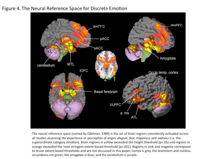
Edelman, G. M. (1989). The remembered present: A biological theory of consciousness. MIT Press. http://www.amazon.com/Remembered-Present-Biological-TheoryConsciousness/dp/046506910X

Eckert, M.A., Menon, V., Walczak, A., Ahlstrom, J., Denslow, S., Horowitz, A., Dubno, J.R. (2009). At the heart of the ventral attention system: The right anterior insula. Human Brain Mapping 30: 2530–2541http://www.ncbi.nlm.nih.gov.ezpprod1.hul.harvard.edu/pmc/articles/PMC2712290/

Ekman, P. & Cordaro, D. (in press). What is meant by calling emotions basic. Emotion Review. Ekman, P. (1999). Basic emotions. In T. Dalgleish & M. J. Powers (Eds.), Handbook of emotion and cognition. New York: John Wiley and Sons LTD.

http://onlinelibrary.wiley.com/book/10.1002/0470013494

Eslinger, P. J., & Damasio, A. R. (1985). Severe disturbance of higher cognition after bilateral frontal lobe ablation: Patient EVR. Neurology 35: 1731.

http://www.neurology.org/cgi/content/abstract/35/12/1731?ijkey=ae100b7b8d72e5bfebd 495730a37f463e8beb641&keytype2=tf_ipsecsha

Etkin, A., & Wager, T. D. (2007). Functional neuroimaging of anxiety: a meta-analysis of emotional processing in PTSD, social anxiety disorder, and specific phobia. The American Journal of Psychiatry 164: 1476-1488.

http://ajp.psychiatryonline.org/cgi/content/abstract/164/10/1476

Ewbank, M. P., Barnard, P. J., Croucher, C. J., Ramponi, C., & Calder, A. J. (2009). The amygdala response to images with impact. Social Cognitive and Affective Neuroscience

4: 127-133.

Fanselow, M. S., & Poulos, A. M. (2005). The neuroscience of mammalian associative learning. Annual Review of Psychology 56: 207-234.

http://arjournals.annualreviews.org/doi/abs/10.1146/annurev..psych.56.091103.070213?u rl_ver=Z39.88-2003&rfr_id=ori:rid:crossref.org&rfr_dat=cr_pub%3Dncbi.nlm.nih.gov

Feinstein, J.S., Adolphs, R., Damasio, A., Tranel, D. (2011). The human amygdala and the induction and experience of fear. Current Biology 21: 34-38.

http://www.ncbi.nlm.nih.gov/pubmed/21167712

Fendt, M., & Fanselow, M. S. (1999). The neuroanatomical and neurochemical basis of conditioned fear. Neuroscience and Biobehavioral Reviews 23: 743–760.

http://psycnet.apa.org/index.cfm?fa=search.displayRecord&uid=1999-05780-008

Ferris, C.F., Stolberg, T., Kulkarni, P., Murugavel, M. Blanchard, R., Blanchard, D.C., et al. (2008). Imaging the neural circuitry and chemical control of aggressive motivation. BMC Neuroscience 9: 1-35. http://www.biomedcentral.com/1471-2202/9/111

Ferry, A., Hespos, S.J., & Waxman, S. (2010). Language facilitates category formation in 3month-old infants. Child Development 81: 472 479.

http://groups.psych.northwestern.edu/infantcognitionlab/FerryHesposWaxman2010.pdf

Fischer, H., Wright, C. I., Whalen, P. J., McInerney, S. C., Shin, L. M., & Rauch, S. L. (2003). Brain habituation during repeated exposure to fearful and neutral faces: a functional MRI study. Brain Research Bulletin 59: 387-392.

http://www.ncbi.nlm.nih.gov/pubmed/12507690

Fontaine, J.R.J., Scherer, K.R., Roesch, E.B., & Ellsworth, P.C. (2007). The world of emotions is not two-dimensional. Psychological Science 18: 1050-1057.

http://pss.sagepub.com/content/18/12/1050.short

Fountoulakis, K.N., Giannakopoulos, P., Kövari, E., & Bouras, C. (2008). Assessing the role of cingulate cortex in bipolar disorder: Neuropathological, structural and functional imaging data. Brain Research Reviews 59: 9-21.

Fox, N. A. (1991). If it's not left, it's right. Electroencephalograph asymmetry and the development of emotion. The American Psychologist 46: 863-872.

http://psycnet.apa.org/index.cfm?fa=buy.optionToBuy&id=1991-32856001&CFID=6336004&CFTOKEN=47588819

Freedman, D. J., Riesenhuber, M., Poggio, T. & Miller, E. K. (2001). Categorical representation of visual stimuli in the primate prefrontal cortex. Science 291: 312–316.

http://www.sciencemag.org/content/291/5502/312.short

Freedman, D. J., Riesenhuber, M., Poggio, T., & Miller, E. K. (2002). Visual categorization and

the primate prefrontal cortex: neurophysiology and behavior. Journal of Neurophysiology

88: 929. http://jn.physiology.org/cgi/content/abstract/88/2/929 Friston, L. (2010). The free energy principle: A unified brain theory? Nature Reviews Neuroscience 11: 127-138

Fugate, J. M. B., Gouzoules, H., & Barrett, L. F. (2010). Reading chimpanzee faces: A test of the structural and conceptual hypotheses: Emotion 10: 544-554. http://www.affectivescience.org/pubs/2010/Fugate_Gouzoules_Barrett_2010CP.pdf

Fusar-Poli, P., Placentino, A., Carletti, F., Landi, P., Allen, P., Surguladze, S., et al. (2009). Functional atlas of emotional faces processing: a voxel-based meta-analysis of 105 functional magnetic resonance imaging studies. Journal of Psychiatry & Neuroscience

34: 418-432. http://www.cma.ca/multimedia/staticContent/HTML/N0/l2/jpn/vol34/issue-6/pdf/pg418.pdf

Fuster, J. M. (2006). The cognit: a network model of cortical representation. International Journal of Psychophysiology 60: 125-132.

http://www.ncbi.nlm.nih.gov/pubmed/16626831

Gendron, M., & Barrett, F., (2009). Reconstructing the past: A century of ideas about emotion in psychology. Emotion Review 1: 316. http://www2.bc.edu/~barretli/pubs/2009/gendronbarrett-2009.pdf

Gendron, M., Lindquist, K.A., Wager, T.D., Bliss-Moreau, E., Kober, H. & Barrett, L.F., (in prep). Seeing with feeling: A meta-analytic review of visual cortex activity in emotion.

Gitelman, D.R., Nobre, A.C., Sonty, S., Parrish, T.B., Mesulam, M.M. (2005). Language network specializations: an analysis with parallel task designs and functional magnetic resonance imaging. Neuroimage 26: 975-985. http://www.ncbi.nlm.nih.gov.ezpprod1.hul.harvard.edu/pubmed/15893473

Glenn, A.L., Raine, A., Schug, R.A. (2009). The neural correlates of moral decision making in psychopathy. Molecular Psychiatry 14: 5-9.

http://www.nature.com/mp/journal/v14/n1/abs/mp2008104a.html

Gotlib, I. H., & Hamilton, J. P. (2008). Neuroimaging and depression: Current status and unresolved issues. Current Directions in Psychological Science 17: 159.

http://psycnet.apa.org/?fa=main.doiLanding&uid=2008-04591-019

Goyer, P. F., Konicki, P. E., & Schulz, S. C. (1994). Brain imaging in personality disorders. In: Ed. K. M. Silk. Biological and Neurobehavioral Studies of Borderline Personality Disorder (pp. 109–125).

Grafman, J., Schwab, K., Warden, D., Pridgen, A., Brown, H. R., & Salazar, A. M. (1996). Frontal lobe injuries and violence a report of the Vietnam Head Injury Study. Neurology

46: 1231–1238. http://www.ncbi.nlm.nih.gov/pubmed/8628458

Greene, J. D., Nystrom, L. E., Engell, A. D., Darley, J. M., & Cohen, J. D. (2004). The neural bases of cognitive conflict and control in moral judgment. Neuron 44: 389–400.

http://www.wjh.harvard.edu/~jgreene/GreeneWJH/Greene-etal-Neuron04.pdf

Gregg, T.R., & Siegel, A. (2001). Brain structures and neurotransmitters regulating aggression in cats: implications for human aggression. Progress in Neuropsychopharacology Biological Psychiatry 25: 91-140. http://www.ncbi.nlm.nih.gov.ezpprod1.hul.harvard.edu/pubmed/11263761

Grezes, J. & Decety, J. (2001). Functional anatomy of execution, mental simulation, observation, and verb generation of actions: A meta-analysis. Human Brain Mapping 12: 1-19. http://www.ncbi.nlm.nih.gov.ezp-prod1.hul.harvard.edu/pubmed/11198101

Grinband, J., Savitsky, J., Wager, T. D., Teichert, T., Ferrera, V. P., & Hirsch, J. (2010). The dorsal medial frontal cortex is sensitive to time on task, not response conflict or error likelihood. Neuroimage.

Gross, J. & Barrett, L.F. (in press). Emotion generation and emotion regulation. One or two depends on your point of view. Emotion Review.

Halgren, E., Walter, R.D., Cherlow, D.G., & Crandall, P.H. (1978). Mental phenomena evoked by electrical stimulation of the human hippocampal formation and amygdala. Brain 101: 83–117. http://www.ncbi.nlm.nih.gov/pubmed/638728

Harenski, C. L., Kim, S. H., & Hamann, S. (2009). Neuroticism and psychopathy predict brain activation during moral and nonmoral emotion regulation. Cognitive, Affective, & Behavioral Neuroscience 9: 1. http://www.ncbi.nlm.nih.gov/pubmed/19246323

Harlow, H. F., & Stagner, R. (1932). Psychology of feelings and emotions. I. Theory of feelings. Psychological Review 39: 570–589. http://psycnet.apa.org/journals/rev/39/6/570.pdf

Harmon-Jones, E., & Allen, J. J. (1998). Anger and frontal brain activity: EEG asymmetry consistent with approach motivation despite negative affective valence. Journal of Personality and Social Psychology 74: 1310–1316. http://psycnet.apa.org/index.cfm?fa=fulltext.journal&jcode=psp&vol=74&issue=5&form at=html&page=1310&expand=1

Harmon-Jones, E., & Sigelman, J. (2001). State anger and prefrontal brain activity: Evidence that insult-related relative left-prefrontal activation is associated with experienced anger and aggression. Journal of Personality and Social Psychology 80: 797–803. http://psycnet.apa.org/journals/psp/80/5/797.html

Harrison, N.A., Gray, M. A., Giarnos, P.J., Critchley, H. G. (2010). The embodiment of emotional feelings in the brain. The Journal of Neuroscience 30: 12878-12884. http://www.ncbi.nlm.nih.gov.ezp-prod1.hul.harvard.edu/pubmed/20861391

Hassabis, D. & Maguire, E.A. (2009). The construction system of the brain. Philosophical Transactions of the Royal Society B 364: 1263-1271. http://rstb.royalsocietypublishing.org/content/364/1521/1263.full

Haxby, J. V., Gobbini, M. I., Furey, M. L., Ishai, A., Schouten, J. L., & Pietrini, P. (2001). Distributed and overlapping representations of faces and objects in ventral temporal cortex. Science 293: 2425. http://www.sciencemag.org/cgi/content/abstract/sci;293/5539/2425

Herry, C., Bach, D. R., Esposito, F., Di Salle, F., Perrig, W. J., Scheffler, K., et al. (2007). Processing of temporal unpredictability in human and animal amygdala. Journal of Neuroscience 27: 5958. http://www.ncbi.nlm.nih.gov/pubmed/17537966

Hitchcock, J. M., & Davis, M. (1986). Lesions of the amygdala, but not of the cerebellum or red nucleus, block conditioned fear as measured with the potentiated startle paradigm. Behavioral Neuroscience 100: 11–22. http://psycnet.apa.org/journals/bne/100/1/11.html

Hitchcock, J. M., & Davis, M. (1987). Fear-potentiated startle using an auditory conditioned stimulus: effect of lesions of the amygdala. Physiology & Behavior 39: 403–408. http://www.ncbi.nlm.nih.gov/pubmed/3575483

Holland, P. C., & Gallagher, M. (1999). Amygdala circuitry in attentional and representational processes. Trends in Cognitive Sciences 3: 65–73. http://www.ncbi.nlm.nih.gov/pubmed/10234229

Hornak, J., Bramham, J., Rolls, E. T., Morris, R. G., O'Doherty, J., Bullock, P. R., et al. (2003). Changes in emotion after circumscribed surgical lesions of the orbitofrontal and cingulate cortices. Brain: A Journal of Neurology 126: 1691-1712. http://www.ncbi.nlm.nih.gov/pubmed/12805109

Horwitz, B. (2005). Integrating neuroscientific data across spatiotemporal scales. Comptes Rendus Biologies 328: 109-118. http://www.ncbi.nlm.nih.gov.ezpprod1.hul.harvard.edu/pubmed/15770997

Hunt, W. A. (1941). Recent developments in the field of emotion. Psychological Bulletin 38: 249–276. http://psycnet.apa.org/?&fa=main.doiLanding&doi=10.1037/h0054615

Izard, C.E. (in press). Forms and functions in emotions: Matters of emotion-cognition interactions. Emotion Review.

Jabbi, M., Bastiaansen, J., & Keysers, C. (2008). A common anterior insula representation of disgust observation, experience and imagination shows divergent functional connectivity pathways. Plosone 3: 1-8.

James, W. (1884). What is an emotion? Mind 9: 188–205. http://www.jstor.org/pss/2246769

James, W. (1890/1998). Principles of Psychology. Chicago, IL: University of Chicago Press.

Jenison, R.L., Rangel, A., Oya, H. Kawasaki, H., Howard, M.A. (2011). Value encoding in single neurons in the human amygdala during decision making. The Journal of Neuroscience 31: 331–338. http://www.ncbi.nlm.nih.gov.ezpprod1.hul.harvard.edu/pubmed/21209219

Jürgens, U. (2009). The Neural Control of Vocalization in Mammals: A Review. Journal of Voice 23: 1-10. http://www.ncbi.nlm.nih.gov/pubmed/8653176

Kamback, M. (1973). The effects of orbital and dorsolateral frontal cortical ablations on ethanol self-selection and emotional behaviors in monkeys. Neuropsychologia 11: 331-335. http://www.ncbi.nlm.nih.gov.ezp-prod1.hul.harvard.edu/pubmed/4209334

Keltner, D. Ekman, P., Gonzaga, G., & Beer, J. (2003). Facial expressions of emotion. In Eds.

R.J. Davidson, K.R. Scherer, H.H. Goldsmith. Handbook of affective sciences (p. 415427). New York Oxford University Press. http://www.amazon.com/Handbook-AffectiveSciences-Science/dp/0195126017

Kennedy, D.P., & Adolphs, R. (2010). Impaired fixation to eyes following amygdala damage arises from abnormal bottom-up attention. Neuropsychologia 48: 3392-3398. http://www.ncbi.nlm.nih.gov/pubmed/20600184

Kensinger, E. A., & Corkin, S. (2004). Two routes to emotional memory: Distinct neural processes for valence and arousal. Proceedings of the National Academy of Sciences 101: 3310–3315. http://www2.bc.edu/elizabeth-kensinger/Kensinger_Corkin_PNAS04.pdf

Kim, J.J., Rison, R.A., Fanselow, M.S., 1993. Effects of amygdala, hippocampus, and periaqueductal gray lesions on short- and long-term contextual fear. Behavioral Neuroscience 107: 1093–1098. http://www.ncbi.nlm.nih.gov.ezpprod1.hul.harvard.edu/pubmed/8136063

Kipps, C.M., Duggins, A.J., McCusker, E.A., & Calder, A.J. (2007). Disgust and happiness recognition correlate with anteroventral insula and amygdala volume respectively in preclinical Huntington’s disease. Journal of Cognitive Neuroscience 19: 1206-1217.

http://www.ncbi.nlm.nih.gov/pubmed/17583995

Knutson, B., Rick, S., Wimmer, G. E., Prelec, D., & Loewenstein, G. (2007). Neural predictors of purchases. Neuron 53: 147-156. http://www.ncbi.nlm.nih.gov/pmc/articles/PMC1876732/

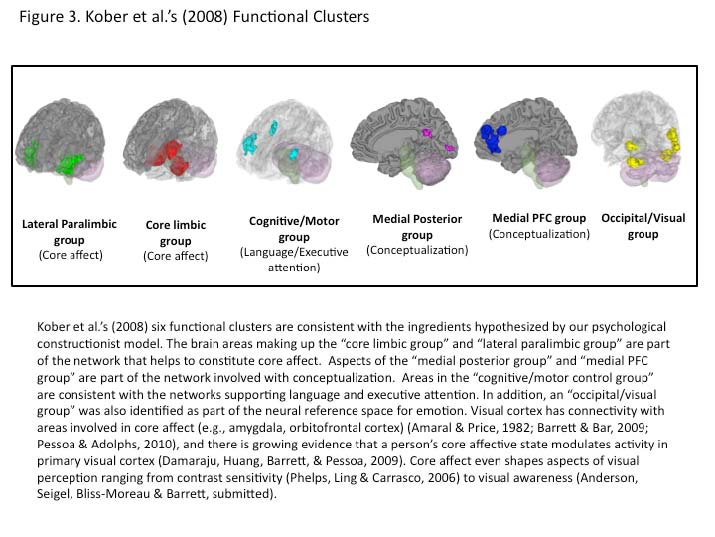
Kober, H., Barrett, L. F., Joseph, J., Bliss-Moreau, E., Lindquist, K., & Wager, T. D. (2008). Functional grouping and cortical-subcortical interactions in emotion: a meta-analysis of neuroimaging studies. NeuroImage 42: 998-1031. http://www.ncbi.nlm.nih.gov/pmc/articles/PMC2752702/

Koenigs M, Young L, Adolphs R, Tranel D, Cushman F, Hauser M, Damasio A. (2007). Damage to the prefrontal cortex increases utilitarian moral judgments. Nature 446: 908

11. http://www.ncbi.nlm.nih.gov/pubmed/17377536

Kopchia, K. L., Altman, H. J., & Commissaris, R. L. (1992). Effects of lesions of the central nucleus of the amygdala on anxiety-like behaviors in the rat. Pharmacology, Biochemistry, and Behavior 43: 453-461. http://www.ncbi.nlm.nih.gov/pubmed/1438482

Krain, A.L., Wilson, A.M., Arbuckle, R., Castellanos, F.X., Milham, M.P. (2006). Distinct neural mechanisms of risk and ambiguity: a meta-analysis of decision-making. Neuroimage 32: 477-484. http://www.ncbi.nlm.nih.gov.ezpprod1.hul.harvard.edu/pubmed/16632383

Kringelbach, M. L., & Rolls, E. T. (2004). The functional neuroanatomy of the human orbitofrontal cortex: Evidence from neuroimaging and neuropsychology. Progress in Neurobiology 72: 341-372. http://www.kringelbach.dk/papers/PN_Kringelbach2004.pdf

LaBar, K. S., Gatenby, J. C., Gore, J. C., LeDoux, J. E., & Phelps, E. A. (1998). Human amygdala activation during conditioned fear acquisition and extinction: a mixed-trial fMRI study. Neuron 20: 937–945. http://www.ncbi.nlm.nih.gov/pubmed/9620698

LaBar, K. S., LeDoux, J. E., Spencer, D. D., & Phelps, E. A. (1995). Impaired fear conditioning following unilateral temporal lobectomy in humans. Journal of Neuroscience 15: 6846. http://www.ncbi.nlm.nih.gov/pubmed/7472442

Laine, C. M., Spitler, K. M., Mosher, C. P., & Gothard, K. M. (2009). Behavioral triggers of skin conductance responses and their neural correlates in the primate amygdala. Journal of Neurophysiology 101: 1749-1754. http://www.ncbi.nlm.nih.gov/pubmed/19144740

Lambon Ralph, M. A., Pobric, G., & Jefferies, E. (2009). Conceptual knowledge is underpinned by the temporal pole bilaterally: convergent evidence from rTMS. Cerebral Cortex 19: 832-838. http://www.ncbi.nlm.nih.gov/pubmed/18678765

Lang, P. J., Davis, M., & Öhman, A. (2000). Fear and anxiety: animal models and human cognitive psychophysiology. Journal of Affective Disorders 61: 137-159. http://www.stanford.edu/~kateri/Becky/PDFs/Lang%202000.pdf

LeDoux, J. (2007). The amygdala. Current Biology 17: R868-874.

LeDoux, J. E., Sakaguchi, A., Iwata, J., & Reis, D. J. (1985). Auditory emotional memories: establishment by projections from the medial geniculate nucleus to the posterior neostriatum and/or dorsal amygdala. Annals of the New York Academy of Sciences 444: 463-464. http://www.ncbi.nlm.nih.gov/pubmed/3860099

LeDoux, J.E., Ciccetti, P., Xagoraris, A., Romanski, L.M. (1990). The lateral amygdaloid nucleus: sensory interface of the amygdala in fear conditioning. Journal of Neuroscience

10: 1062-1069. http://www.ncbi.nlm.nih.gov/pubmed/2329367

LeDoux, J.E., Sakaguchi, A. & Reis, D.J (1983). Strain differences in fear between spontaneously hypertensive and normotensive rats. Brain Research 277: 137-143.

http://www.ncbi.nlm.nih.gov/pubmed/6640286

Lindquist, K. A., & Barrett, L. F. (2008). Constructing emotion: The experience of fear as a conceptual act. Psychological Science 19: 898.

http://www2.bc.edu/~lindqukr/docs/Lindquist&Barrett2008.pdf

Lindquist, K. A., Barrett, L. F., Bliss-Moreau, E., & Russell, J. A. (2006). Language and the perception of emotion. Emotion 6: 125-138.

http://nmr.mgh.harvard.edu/~lindqukr/publications.html

Lovick, T.A., 1992. Inhibitory modulation of the cardiovascular defence response by the ventrolateral periaqueductal grey matter in rats. Experimental Brain Research 89: 133–

139. http://www.springerlink.com/content/h18327q02g311765/

Machado, C.J., Bachevalier, J. (2006). The impact of selective amygdala, orbital frontal cortex, or hippocampal formation lesions on established social relationships in rhesus monkeys (Macaca mulatta). Behavioral Neuroscience 120:761-786.

http://www.ncbi.nlm.nih.gov/pubmed/16893284

MacLean, P.D., & Newman, J.D., (1988). Role of midline frontolimbic cortex in production of the isolation call of squirrel monkeys. Brain Research 450: 111-123.

http://www.ncbi.nlm.nih.gov.ezp-prod1.hul.harvard.edu/pubmed/3401705

Mandler, G. (1975). Mind and emotion. New York: Wiley. Mandler, G. (1990). William James and the construction of emotion. Psychological Science 1: 179-180. http://www.jstor.org/stable/40062633

Mansouri, F.A., Tanaka, K., Buckley, M.J. (2009). Conflict-induced behavioral adjustment: A clue to the executive functions of prefrontal cortex. Nature Reviews Neuroscience 10: 141-152. http://www.ncbi.nlm.nih.gov.ezp-prod1.hul.harvard.edu/pubmed/19153577

Maskati, A.A.A & Zbrozyna, A.W. (1989). Cardiovascular and motor components of the defense reaction elicited in rats by electrical and chemical stimulation in amygdala. Journal of the Autonomic Nervous System 28: 127-132. http://www.ncbi.nlm.nih.gov.ezpprod1.hul.harvard.edu/pubmed/2625501

Mason, W. A., Capitanio, J. P., Machado, C. J., Mendoza, S. P., & Amaral, D. G. (2006). Amygdalectomy and responsiveness to novelty in rhesus monkeys (Macaca mulatta): generality and individual consistency of effects. Emotion 6: 73-81. doi: 10.1037/15283542.6.1.73.

http://psycnet.apa.org/index.cfm?fa=fulltext.journal&jcode=emo&vol=6&issue=1&form at=html&page=73&expand=1

Matynh, P.W., (1983). Connections of midbrain periaqueductal gray in the monkey. I. Ascending efferent projections. Journal of Neurophysiology 49: 567-581.

http://www.ncbi.nlm.nih.gov.ezp-prod1.hul.harvard.edu/pubmed/6300350

Mauss, I. B., & Robinson, M. D. (2009). Measures of emotion: A review. Cognition & Emotion

23: 209. http://www.ncbi.nlm.nih.gov/pmc/articles/PMC2756702/

Mayberg, H. S., Lozano, A. M., Voon, V., McNeely, H. E., Seminowicz, D., Hamani, C., et al. (2005). Deep brain stimulation for treatment-resistant depression. Neuron 45:651–660.

http://www.ncbi.nlm.nih.gov/pubmed/15748841

McDermott, K.B., Szpunar, K.K., & Christ, S.E. (2009). Laboratory-based and autobiographical retrieval tasks differ substantially in their neural substrates. Neuropsychologia 47: 22902298. http://memory.wustl.edu/McDermott_Lab/Publications.html

McIntosh, A. R., Bookstein, F. L., Haxby, J. V., & Grady, C. L. (1996). Spatial pattern analysis of functional brain images using partial least squares. Neuroimage 3: 143–157.

http://www.ncbi.nlm.nih.gov/pubmed/9345485

McIntosh, A. R. (2004). Contexts and catalysts: A resolution of the location and integration of function in the brain. Neuroinformatics 2: 175-181 http://www.springerlink.com.ezpprod1.hul.harvard.edu/content/k12p31883927q237/

Mendes, W. B., Blascovich, J., Hunter, S. B., Lickel, B., & Jost, J. T. (2007). Threatened by the unexpected: Physiological responses during social interactions with expectancy-violating partners. Journal of Personality and Social Psychology 92: 698.

http://psycnet.apa.org/journals/psp/92/4/698.html

Mesulam, M.M. & Mufson, E.J. (1982). Insula of the old world monkey. 111: Efferent cortical output and comments on function. The Journal of Comparative Neurology 212: 38-52.

http://www3.interscience.wiley.com/journal/109687236/abstract?CRETRY=1&SRETRY =0

Milders, M., Crawford, J. R., Lamb, A., & Simpson, S. A. (2003). Differential deficits in expression recognition in gene-carriers and patients with Huntington's disease. Neuropsychologia 41: 1484-1492. http://www.ncbi.nlm.nih.gov/pubmed/12849766

Milham, M.P., Banich, M.T., Webb, A., Barad, V., Cohen, N.J., Wszalek, T., Kramer, A.F., (2001). The relative involvement of anterior cingulate and prefrontal cortex in attentional control depends on nature of conflict. Brain Research. Cognitive Brain Research 12: 467

473. http://www.ncbi.nlm.nih.gov.ezp-prod1.hul.harvard.edu/pubmed/11689307

Miller, E. K. (2000). The prefrontal cortex and cognitive control. Nature Reviews Neuroscience

1: 59-65. http://www.doc.ic.ac.uk/~xh1/Referece/Visual-search/neural-cognitivecontrl.pdf

Miller, E. K., & Cohen, J. D. (2001). An integrative theory of prefrontal cortex function. Annual Review of Neuroscience 24: 167-202.

http://arjournals.annualreviews.org/doi/abs/10.1146%2Fannurev.neuro.24.1.167

Miller, E. K., Freedman, D. J., & Wallis, J. D. (2002). The prefrontal cortex: categories, concepts and cognition. Philosophical Transactions: Biological Sciences 357: 1123–1136.

http://rstb.royalsocietypublishing.org/content/357/1424/1123.full.pdf

Missilin, R., & Ropartz, P. (1981). Effects of lateral amygdala lesions on the responses to novelty in mice. Behavioural Processes 6: 329-336.

Mitchell, I. J., Heims, H., Neville, E. A., & Rickards, H. (2005). Huntington's disease patients show impaired perception of disgust in the gustatory and olfactory modalities. Journal of Neuropsychiatry and Clinical Neurosciences 17: 119.

http://www.ncbi.nlm.nih.gov/pubmed/15746492

Mitchell, J.P. (2009). Inferences about other people’s minds. Philosophical Transactions of the Royal Society B 364: 1309-1316.

http://www.wjh.harvard.edu/~scanlab/papers/2009_predictions_PhilTrans.pdf

Mobbs, D., Petrovic, P., Marchant, J.L., Hassabis, D., Weiskopf, N., Seymour, B., et al., (2007). When fear is near: threat imminence elicits prefrontal periaqueductal gray shifts in humans. Science 317: 1079. http://www.ncbi.nlm.nih.gov/pmc/articles/PMC2648508/

Moriguchi, Y. Negreira, A., Weirerich, M., Dautoff, R., Dickerson, B.C., Wright, C.I., Barrett, L.

F. (2010). Differential hemodynamic response in affective circuitry with aging: novely, valence and arousal. Journal of Cognitive Neuroscience.

http://www.mitpressjournals.org.ezpprod1.hul.harvard.edu/doi/pdf/10.1162/jocn.2010.21527

Moulton, S.T., & Kosslyn, S. (2009). Imagining predictions: Mental imagery as mental emulation. Philosophical Transactions of the Royal Society B 364: 1273-1280. http://rstb.royalsocietypublishing.org/content/364/1521/1273.full

Moyer, K.E., Kinds of aggression and their physiological basis, Communications in Behavioral Biology, A2 (1968) 65-87.

Murphy, F. C., Nimmo-Smith, I., & Lawrence, A. D. (2003). Functional neuroanatomy of emotions: A meta-analysis. Cognitive, Affective & Behavioral Neuroscience 3: 207-233. http://www.ncbi.nlm.nih.gov/pubmed/14672157

Nachman, M., & Ashe, J. H. (1974). Effects of basolateral amygdala lesions on neophobia, learned taste aversions, and sodium appetite in rats. Journal of Comparative and Physiological Psychology 87: 622-643. http://psycnet.apa.org/journals/com/87/4/622.pdf

Nelson, J.K., Reuter-Lorenz, P.A., Sylvester, C.C., Jonides, J. & Smith, E.E. (2003). Dissociable neural mechanisms underlying response-based and familiarity-based conflict in working memory. Proceedings of the National Academy of Sciences 100: 11171–11175. http://www.ncbi.nlm.nih.gov.ezp-prod1.hul.harvard.edu/pubmed/12958206

Newman, S. W. (1999). The medial extended amygdala in male reproductive behavior. A node in the mammalian social behavior network. Annals of the New York Academy of Sciences 877: 242-57. http://www.ncbi.nlm.nih.gov/sites/entrez

Northoff, (2006). Is emotion regulation self-regulation? Trends in Cognitive Sciences 9: 408

409. http://dept.psych.columbia.edu/~kochsner/pdf/Northoff_Tics_comment.pdf

Numan, M. (2007). Motivational systems and the neural circuitry of maternal behavior in the rat. Developmental Psychobiology 49: 12-21. http://www.ncbi.nlm.nih.gov/pubmed/17186513

Ochsner, K. N., Ray, R. D., Cooper, J. C., Robertson, E. R., Chopra, S., Gabrieli, J. D. E., et al. (2004). For better or for worse: neural systems supporting the cognitive down- and up-regulation of negative emotion. NeuroImage 23:483-499. http://www.ncbi.nlm.nih.gov/pubmed/15488398

Ochsner, K. N., Knierim, K., Ludlow, D., Hanelin, J., Ramachandran, T. & Mackey, S. (2004). Reflecting upon feelings: An fMRI study of neural systems supporting the attribution of emotion to self and other. Journal of Cognitive Neuroscience 16: 1748-1772. http://dept.psych.columbia.edu/~kochsner/pdf/Ochsner_Reflect_Feelings.pdf

Öhman, A. (2009). Of snakes and faces: An evolutionary perspective on the psychology of fear. Scandinavian Journal of Psychology 50: 543-552. http://www.ncbi.nlm.nih.gov.ezpprod1.hul.harvard.edu/pubmed/19930253

Öngür, D., & Price, J. L. (1998). Prefrontal cortical projections to the hypothalamus in macaque monkeys. Journal of Comparative Neurology 30: 480-505. http://www3.interscience.wiley.com/journal/5005316/abstract

Öngür, D., & Price, J. L. (2000). The organization of networks within the orbital and medial prefrontal cortex of rats, monkeys and humans. Cerebral Cortex 10: 206.

http://cercor.oxfordjournals.org/cgi/content/abstract/10/3/206

Ortigue, S., Grafton, S. T., & Bianchi-Demicheli, F. (2007). Correlation between insula activation and self-reported quality of orgasm in women. NeuroImage 37: 551-560. http://www.ncbi.nlm.nih.gov/pubmed/17601749

Ortony A, & Turner, T.J. (1990). What’s basic about basic emotions? Psychological Review 97:315–331.http://web.ebscohost.com.ezpprod1.hul.harvard.edu/ehost/detail?hid=10&sid=79dcf313-fa01-4d4c-aff0542b9341511a%40sessionmgr13&vid=4&bdata=JnNpdGU9ZWhvc3QtbGl2ZSZzY29w ZT1zaXRl#db=psyh&AN=1990-27526-001

Owen, A.M., McMillan, K., Laird, A.R., Bullmore, E. (2005). N-Back working memory paradigm: A meta-analysis of normative functional neuroimaging studies. Human Brain Mapping 25: 46 –59. http://www.ncbi.nlm.nih.gov.ezpprod1.hul.harvard.edu/pubmed/15846822

Panksepp, J. & Watt, D. (in press). What is basic about basic emotions? Lasting lessons from affective neuroscience. Emotion Review.

Panksepp, J. (1998). Affective neuroscience: The foundations of human and animal emotions. New York: Oxford University Press.

Panksepp, J. (2007). Neurologizing the psychology of affects: How appraisal-based constructivism and basic emotion theory can coexist. Perspectives on Psychological Science 2: 281. http://www3.interscience.wiley.com/journal/118509152/abstract

Paton, J.P., Belova, M.A., Morrison, S.E. & Salzman, C.D. (2006). The primate amygdala represents the positive and negative value of visual stimuli during learning. Nature 16: 865-870. http://www.ncbi.nlm.nih.gov/pmc/articles/PMC2396495/

Paus, T. (2001). Primate anterior cingulate cortex: Where motor control, drive and cognition interface. Nature Reviews Neuroscience 2: 417–424. http://www.oise.utoronto.ca/research/brainwaves/phpwebsite/files/uplink/Paus_2001.pdf

Penfield, W., & Falk, M.E. (1955). The insula. Further observations on its function. Brain 78: 445-470. http://brain.oxfordjournals.org/content/78/4/445.extract

Pessoa, L. (2010). Emotion and cognition and the amygdala: From “what is it?” to “what's to be done?”. Neuropsychologia 48: 3416-3429. http://www.indiana.edu/%7Elceiub/publications_files/Pessoa_Neuropsychologia_2010.p df

Pessoa, L. & Adolphs, R. (2010). Emotion processing and the amygdala: from a ‘low road’ to ‘many roads’ of evaluating biological significance. Nature Reviews Neuroscience 11: 773-783. http://www.indiana.edu/%7Elceiub/publications_files/Pessoa_Adolphs_NRN_2010_with _suppl.pdf

Pessoa, L. (2008). On the relationship between emotion and cognition. Nature Reviews Neuroscience 9: 148-158.

Petrides, M. (2005). Lateral prefrontal cortex: architectonic and functional organization.

Philosophical Transactions of the Royla Society of London: B Biological Sciences 360:781-95. http://www.ncbi.nlm.nih.gov.ezp-prod1.hul.harvard.edu/pubmed/15937012

Petrides, M. (2007). The orbitofrontal cortex: novelty, deviation from expectation, and memory. Annals of the New York Academy of Sciences 1121: 33-53. http://www.ncbi.nlm.nih.gov/pubmed/17872393

Phan, K. L., Wager, T., Taylor, S. F., & Liberzon, I. (2002). Functional neuroanatomy of emotion: a meta-analysis of emotion activation studies in PET and fMRI. NeuroImage

16: 331-348. http://www.ncbi.nlm.nih.gov/pubmed/12030820

Pobric, G., Jefferies, E., & Ralph, M. A. L. (2007). Anterior temporal lobes mediate semantic representation: mimicking semantic dementia by using rTMS in normal participants. Proceedings of the National Academy of Science 104: 20137-20141. http://www.pnas.org/content/104/50/20137.abstract

Poldrack, R.A., Halchenko, Y.O., Hanson, S.J. (2009). Decoding the large-scale structure of brain function by classifying mental States across individuals. Psychological Science 20: 1364-1372. http://www.ncbi.nlm.nih.gov.ezp-prod1.hul.harvard.edu/pubmed/19883493

Price, C.J., & Friston, K.J. (2005). Functional ontologies for cognition: The systematic definition of structure and function. Cognitive Neuropsychology 22: 262-275. http://www.ncbi.nlm.nih.gov.ezp-prod1.hul.harvard.edu/pubmed/21038249

Raichle, M. E., MacLeod, A. M., Snyder, A. Z., Powers, W. J., Gusnard, D. A., & Shulman, G.

L. (2001). A default mode of brain function. Proceedings of the National Academy of Sciences 98: 676-682. http://www.pnas.org/content/98/2/676.long

Raine, A., Lencz, T., Bihrle, S., LaCasse, L., & Colletti, P. (2000). Reduced prefrontal gray matter volume and reduced autonomic activity in antisocial personality disorder. Archives of General Psychiatry 57: 119-127. http://www.ncbi.nlm.nih.gov/pubmed/10665614

Raleigh, M. J., Steklis, H. D., Ervin, F. R., Kling, A. S., & McGuire, M. T. (1979). The effects of orbitofrontal lesions on the aggressive behavior of vervet monkeys (Cercopithecus aethiops sabaeus). Experimental Neurology 66: 158-168. http://www.ncbi.nlm.nih.gov/pubmed/113236

Rempel-Clower, N.L., & Barbas H. (1998). Topographic organization of connections between the hypothalamus and prefrontal cortex in the rhesus monkey. Journal of Comparative Neurology 398: 393-419. http://www.ncbi.nlm.nih.gov/pubmed/9714151

Rankin, K.P. Gorno-Tempini, M.L., Allison, S.C., Stanley, C.M., Glenn, S., Weiner, M.W., Miller, B.L. (2006). Structural anatomy of empathy in neurodegenerative disease. Brain

129: 2945-2956.

Reynolds, S. M., & Berridge, K. C. (2002). Positive and negative motivation in nucleus accumbens shell: Bivalent rostrocaudal gradients for GABA-elicited eating, taste "liking"/"disliking" reactions, place preference/avoidance, and fear. The Journal of Neuroscience 22: 7308-7320. http://www.jneurosci.org/cgi/content/abstract/22/16/7308 Reynolds, S. M., & Berridge, K. C. (2003). Glutamate motivational ensembles in nucleus accumbens: Rostrocaudal shell gradients of fear and feeding. European Journal of Neuroscience 17: 2187–2200. http://www.ncbi.nlm.nih.gov/pubmed/12786986

Reynolds, S. M., & Berridge, K. C. (2008). Emotional environments retune the valence of appetitive versus fearful functions in nucleus accumbens. Nature Neuroscience 11: 423

425. http://www.ncbi.nlm.nih.gov/pmc/articles/PMC2717027/

Roberson, D., & Davidoff, J. (2000). The categorical perception of color and facial expressions: The effect of verbal interference. Memory & Cognition 28: 977–986. http://www.ncbi.nlm.nih.gov/pubmed/11105523

Roberson, D., Damjanovic, L. & Pilling, M. (2007).Categorical perception of facial expressions: Evidence for a 'category adjustment' model. Memory & Cognition 35: 1814-1829.

Rogers, T. T., Lambon Ralph, M. A., Garrard, P., Bozeat, S., McClelland, J. L., Hodges, J. R., et al. (2004). Structure and deterioration of semantic memory: A neuropsychological and computational investigation. Psychological Review 111: 205-235. http://charlotte.cnbc.cmu.edu/ibsc/papers/RogersETAL04_PR.pdf

Rolls E.T., Critchley, H.D., Mason, R., Wakeman EA. (1996). Orbitofrontal cortex neurons: role in olfactory and visual association learning. Journal of Neurophysiology 75: 1970-1981. http://www.ncbi.nlm.nih.gov/pubmed/8734596

Rolls, E. (1999). The function of the orbitofrontal cortex. Neurocase 5: 301-312.

http://www.oxcns.org/papers/357_Rolls04.pdf

Rolls, E.T., Hornak, J., Wade, D., & McGrath J. (1994). Emotion-related learning in patients with social and emotional changes associated with frontal lobe damage. Journal of Neurology, Neurosurgery and Psychiatry 57:1518-1524. http://www.ncbi.nlm.nih.gov/pubmed/7798983

Roseman, I.J. (1984). Cognitive determinants of emotion: A structural theory. Review of Personality & Social Psychology 5: 11-36. http://web.ebscohost.com.ezpprod1.hul.harvard.edu/ehost/results?hid=10&sid=79dcf313-fa01-4d4c-aff0542b9341511a%40sessionmgr13&vid=5&bquery=%28AU+%28Roseman%29%29+and +%28DT+1984%29&bdata=JmRiPXBzeWgmdHlwZT0xJnNpdGU9ZWhvc3QtbGl2ZS ZzY29wZT1zaXRl

Rosen, H. J., Pace-Savitsky, K., Perry, R. J., Kramer, J. H., Miller, B. L., & Levenson, R. W. (2004). Recognition of emotion in the frontal and temporal variants of frontotemporal dementia. Dementia and Geriatric Cognitive Disorders 17: 277-281. http://www.ncbi.nlm.nih.gov/pubmed/15178936

Rosen, J.B. & Davis, M. (1988). Enhancement of acoustic startle by electrical stimulation of the amygdala. Behavioral Neuroscience 102: 195-202. http://www.sciencedirect.com/science?_ob=ArticleURL&_udi=B6WY4-46X46NT4F&_user=10&_coverDate=04%2F30%2F1988&_rdoc=1&_fmt=high&_orig=search&_ origin=search&_sort=d&_docanchor=&view=c&_searchStrId=1509840975&_rerunOrigi n=scholar.google&_acct=C000050221&_version=1&_urlVersion=0&_userid=10&md5= e681bf616267abecaf65243450732d0d&searchtype=a

Rozin, P., Haidt, J., & McCaule, C. (2008). Disgust. In: The Handbook of Emotions. Eds. M. Lewis, J.M. Haviland-Jones and L.F. Barrett. (3rd ed. pp. 757-776). New York: Guilford.

Rozin, P., Haidt, J., & McCauley, C. (2000). Disgust. In: Eds. M. Lewis and J.M. Havilland-Jones. Handbook of emotions (2nd ed., pp. 637-653). New York: Guilford.

Rushworth, M.F., Buckley, M.J., Behrens, T.E., Walton, M.E., Bannerman, D.M. (2007). Functional organization of the medial frontal cortex. Current Opinion in Neurobiology

17: 220-227. http://www.ncbi.nlm.nih.gov.ezp-prod1.hul.harvard.edu/pubmed/17350820

Russell, J. A. (2003). Core affect and the psychological construction of emotion. Psychological Review 110: 145–172.

http://psycnet.apa.org/index.cfm?fa=fulltext.journal&jcode=rev&vol=110&issue=1&for mat=html&page=145&expand=1

Russell, J.A., Bachorowski, J., & Fernandez-Dols, J.M. (2003). Facial and vocal expressions of emotion. Annual Review of Psychology 54: 329-349.

https://www2.bc.edu/~russeljm/publications/annurev.psych.2003.pdf

Russell, J. A., & Barrett, L. F. (1999). Core affect, prototypical emotional episodes, and other things called emotion: Dissecting the elephant. Journal of Personality and Social Psychology 76: 805-819. http://psycnet.apa.org/journals/psp/76/5/805.html

Russell, J.A., & Bullock, M. (1986). On the dimensions preschoolers use to interpret facial expressions of emotion. Developmental Psychology 22: 97-102.

http://www2.bc.edu/~russeljm/

Sander, D., Grafman, J., & Zalla, T. (2003). The human amygdala: An evolved system for relevance detection. Reviews in the Neurosciences 14: 303–316.

http://www.affective-sciences.org/system/files/Sander_etal_RevNeuro.PDF

Sander, D., Grandjean, D, Kaiser, S., Wehrle, T., & Scherer, K.R. (2007). Interaction effects of perceived gaze direction and dynamic facial expression: Evidence for appraisal theories of emotion. European Journal of Cognitive Psychology 19: 470-480. http://www.lemanicneuroscience.ch/PENSTrainingCenter/articles/dsdg_ejcp06.pdf

Saver, J. L., & Damasio, A. R. (1991). Preserved access and processing of social knowledge in a patient with acquired sociopathy due to ventromedial frontal damage. Neuropsychologia

29: 1241-1249. http://www.ncbi.nlm.nih.gov/pubmed/1791934

Saxe, R. & Kanwisher, N. (2003). People thinking about people: The role of the temporoparietal junction in “theory of mind.” Neuroimage 19: 1835-1842.

http://web.mit.edu/bcs/nklab/media/pdfs/SaxeKanwisherNeuroImage03.pdf

Schachter, S., & Singer, J. (1962). Cognitive, social, and physiological determinants of an emotional state. Psychological Review 69: 379-399.

http://www.ncbi.nlm.nih.gov/pubmed/14497895

Schacter, D. L., Addis, D. R., & Buckner, R. L. (2007). Remembering the past to imagine the future: the prospective brain. Nature Reviews Neuroscience 8: 657-661.

http://www.nature.com/nrn/journal/v8/n9/abs/nrn2213.html

Shackman, A.J., Salomons, T.V., Slagter, H.A., Fox, A.S., Winter, J.J., Davidson, R.J. (2011). The integration of negative affect, pain, and cognitive control in the cingulate cortex. Nature Reviews Neuroscience 12: 154-167. http://www.ncbi.nlm.nih.gov.ezpprod1.hul.harvard.edu/pubmed/21331082

Scherer, K.R., & Ellsworth, P.C. (2003). Appraisal processes in emotion. In R.J. Davidson, K.R. Scherer, & H. Goldsmith (Eds.), Handbook of affective sciences (pp. 572–589). New York: Oxford University Press. http://www.amazon.com/Handbook-Affective-SciencesScience/dp/0195126017

Schienle, A., & Schäfer, A. (2009). In search of specificity: functional MRI in the study of emotional experience. International Journal of Psychophysiology 73: 22-26.

http://www.ncbi.nlm.nih.gov/pubmed/19530276

Schnur, T. T., Schwartz, M. F., Kimberg, D. Y., Hirshorn, E., Coslett, H. B., & Thompson-Schill, S. L. (2009). Localizing interference during naming: Convergent neuroimaging and neuropsychological evidence for the function of Broca's area. Proceedings of the National Academy of Sciences 106: 322-327. http://nihongo.j

talk.com/kanji/?link=http://www.pnas.org/content/106/1/322.full

Schwartz, C. E., Wright, C. I., Shin, L. M., Kagan, J., Whalen, P. J., McMullin, K. G., et al. (2003). Differential amygdalar response to novel versus newly familiar neutral faces: a functional MRI probe developed for studying inhibited temperament. Biological Psychiatry 53: 854-862. http://www.whalenlab.info/Publications%20Page/PDFs/Schwartz_2003.pdf

Shultz, W. (2006). Behavioral theories and the neurophysiology of reward. Annual Review of Psychology 57: 87-115. http://www.ncbi.nlm.nih.gov.ezpprod1.hul.harvard.edu/pubmed/16318590

Scott, S. K., Young, A. W., Calder, A. J., Hellawell, D. J., Aggleton, J. P., & Johnson, M. (1997). Impaired auditory recognition of fear and anger following bilateral amygdala lesions. Nature 385: 254–257. http://www.cf.ac.uk/psych/home2/papers/aggleton/Nature%20-%20385%20-%20254257.pdf

Seeley, W. W., Menon, V., Schatzberg, A. F., Keller, J., Glover, G. H., Kenna, H., et al. (2007). Dissociable intrinsic connectivity networks for salience processing and executive control. Journal of Neuroscience 27: 2349. http://neuro.cjb.net/cgi/content/abstract/27/9/2349

Siegel, A., Edinger, H., & Dotto, M. (1975). Effects of electrical stimulation of the lateral aspect of the prefrontal cortex upon attack behavior in cats. Brain Research 93: 473-484. http://www.sciencedirect.com/science?_ob=ArticleURL&_udi=B6SYR-4840TTX32G&_user=10&_coverDate=08%2F15%2F1975&_rdoc=1&_fmt=high&_orig=search& _origin=search&_sort=d&_docanchor=&view=c&_searchStrId=1509842536&_rerunOri gin=scholar.google&_acct=C000050221&_version=1&_urlVersion=0&_userid=10&md 5=847c9d2d236f8c1c60a7f4b0674543ee&searchtype=a

Siegel, A., Edinger, H., & Lowenthal, H. (1974). Effects of electrical stimulation of the medial aspect of the prefrontal cortex upon attack behavior in cats. Brain Research 66: 467-479. http://www.sciencedirect.com/science?_ob=ArticleURL&_udi=B6SYR-484DBF6D8&_user=10&_coverDate=02%2F08%2F1974&_rdoc=1&_fmt=high&_orig=search&_ origin=search&_sort=d&_docanchor=&view=c&_searchStrId=1509841573&_rerunOrigi n=scholar.google&_acct=C000050221&_version=1&_urlVersion=0&_userid=10&md5= 35b6ce6d6686d719eef9f30bb51da726&searchtype=a

Simmons, W.K., Hamann, S.B., Harenski, C.N., Hu, X.P., & Barsalou, L.W. (2008). fMRI evidence for word association and situated simulation in conceptual processing. Journal of Physiology – Paris 102: 106-119. http://psychology.emory.edu/cognition/barsalou/onlinepapers.html

Smith S.M., Fox, P.T., Miller, K.L., Glahn, D.C., Fox, P.M., Mackay, C.E., et al. (2009). Correspondence of the brain’s functional architecture during activation and rest. Proceedings of the National Academy of Sciences 106: 13040–13045. http://www.ncbi.nlm.nih.gov.ezp-prod1.hul.harvard.edu/pubmed

Spinks, J. A., Blowers, G. H., & Shek, D. T. (1985). The role of the orienting response in the anticipation of information: A skin conductance response study. Psychophysiology 22: 385-394. http://www.ncbi.nlm.nih.gov/pubmed/4023149

Sprengelmeyer, R. (2007). The neurology of disgust. Brain 130: 1715-1717.

http://www.ncbi.nlm.nih.gov/pubmed/17550907

Sprengelmeyer, R., Young, A. W., Schroeder, U., Grossenbacher, P. G., Federlein, J., Büttner, T., et al. (1999). Knowing no fear. Proceedings of the Royal Society B: Biological Sciences 266: 2451. http://www.ncbi.nlm.nih.gov/pmc/articles/PMC1690477/

Sprengelmeyer, R., Young, A.W., Calder, A.J., Karnat, A., Lange, H., Homberg, V., Perrett, D.I., Rowland, D. (1996). Loss of disgust. Perception of faces and emotions in Huntington's disease. Brain 119: 1647-1665. http://www.ncbi.nlm.nih.gov.ezpprod1.hul.harvard.edu/pubmed?term=Sprengelmeyer%20et%20al.%201996

Sprengelmeyer R, Rausch M, Eysel UT, Przuntek H. (1998). Neural structures associated with recognition of facial expressions of basic emotions. Proceedings of the Royal Society: Biological Sciences 265:1927-1931. http://www.ncbi.nlm.nih.gov.ezpprod1.hul.harvard.edu/pubmed/9821359

Spreng, R.N., & Mar, R.A. (in press). I remember you: A role for memory in social cognition and the functional neuroanatomy of their interaction. Brain Research.

Sutton, S. K., & Davidson, R. J. (1997). Prefrontal brain asymmetry: A biological substrate of the behavioral approach and inhibition systems. Psychological Science 8: 204–210. http://www.jstor.org/stable/40063179?cookieSet=1

Suvak, M. K., & Barrett, L. F. (2011). Considering PTSD from the perspective of brain processes: A psychological construction analysis. Journal of Traumatic Stress 24: 3-24. http://www.affective-science.org/pubs/2011/suvak-barrett-2011.pdf

Suzuki, A., Hoshino, T., Shigemasu, K., & Kawamura, M. (2006). Disgust-specific impairment of facial expression recognition in Parkinson's disease. Brain 129: 707. http://brain.oxfordjournals.org/cgi/content/abstract/129/3/707

Svoboda, E., McKinnon, M.C., Levine, B. (2006). The functional neuroanatomy of autobiographical memory: A meta-analysis. Neuropsychologia 44: 2189–2208. http://www.ncbi.nlm.nih.gov.ezp-prod1.hul.harvard.edu/pubmed/16806314

Tomkins, S. S. (1962), Affect Imagery Consciousness: The Positive Affects (Vol. 1). New York: Springer.

Tomkins, S. S. (1963), Affect Imagery Consciousness: The Negative Affects (Vol. 2). New York: Springer.

Tsakiris, M., Hesse, M. D., Boy, C., Haggard, P., & Fink, G. R. (2007). Neural Signatures of Body Ownership: A Sensory Network for Bodily Self-Consciousness. Cerebral Cortex

17: 2235-2244. http://cercor.oxfordjournals.org/cgi/content/full/17/10/2235

Tsuchiya, N., Moradi, F., Felsen, C., Yamazaki, M., & Adolphs, R. (2009). Intact rapid detection of fearful faces in the absence of the amygdala. Nature Neuroscience 12: 1224-1225. http://www.ncbi.nlm.nih.gov/pmc/articles/PMC2756300/

Urry, H. L., van Reekum, C. M., Johnstone, T., Kalin, N. H., Thurow, M. E., Schaefer, H. S., et al. (2006). Amygdala and ventromedial prefrontal cortex are inversely coupled during regulation of negative affect and predict the diurnal pattern of cortisol secretion among older adults. The Journal of Neuroscience 26: 4415-4425. http://neuro.cjb.net/cgi/content/abstract/26/16/4415

Van der Horst, V.G., Holstege, G., 1998. Sensory and motor components of reproductive behavior: pathways and plasticity. Behavior and Brain Research 92: 157–167. http://www.ncbi.nlm.nih.gov.ezp-prod1.hul.harvard.edu/pubmed

Van Snellenberg, J.X. & Wager, T.D. (in press). Cognitive and motivational functions of the human prefrontal cortex. In E. Goldberg and D. Bougakov (Eds). Luria’s Legacy in the 21st Century. New York: Oxford. http://www.columbia.edu/cu/psychology/tor/Papers/luria_vans_wager_sub.pdf

Vazdarjanova, A., & McGaugh, J. L. (1998). Basolateral amygdala is not critical for cognitive memory of contextual fear conditioning. Proceedings of the National Academy of Sciences 95: 15003-15007. http://www.pnas.org/content/95/25/15003.abstract

Vigneau, M., Beaucousin, V., Hervé, P. Y., Duffau, H., Crivello, F., Houdé, O., et al. (2006). Meta-analyzing left hemisphere language areas: phonology, semantics, and sentence processing. NeuroImage 30: 1414-1432. http://www.ncbi.nlm.nih.gov/pubmed/16413796

Vincent, J. L., Snyder, A. Z., Fox, M. D., Shannon, B. J., Andrews, J. R., Raichle, M. E., et al. (2006). Coherent spontaneous activity identifies a hippocampal-parietal memory network. Journal of Neurophysiology 96: 3517-3531. http://jn.physiology.org/cgi/content/abstract/96/6/3517

Vogt, B. A., Nimchinsky, E. A., Vogt, L. J., & Hof, P. R. (1995). Human cingulate cortex: Surface features, flat maps and cytoarchitecture. The Journal of Comparative Neurology

359: 490-506. http://onlinelibrary.wiley.com/doi/10.1002/cne.903590310/abstract

Vogt, B.A., Berger, G.R., & Derbyshire, S.W. (2003). Structural and functional dichotomy of human midcingulate cortex. European Journal of Neuroscience 18: 3134-3144. http://www.ncbi.nlm.nih.gov.ezp-prod1.hul.harvard.edu/pubmed/14656310

von dem Hagen, E.A., Beaver, J.D., Ewbank, M.P., Keane, J., Passamonti, L., Lawrence, A.D., Calder, A.J. (2009). Leaving a bad taste in your mouth but not in my insula. Social Cognitive Affective Neuroscience 4: 379-86. http://www.ncbi.nlm.nih.gov/pubmed/19505971

Vuilleumier, P. (2005). How brains beware: neural mechanisms of emotional attention. Trends in Cognitive Sciences 12: 585-594. http://www.ncbi.nlm.nih.gov.ezpprod1.hul.harvard.edu/pubmed/16289871

Vuilleumier, P., & Driver, J. (2007). Modulation of visual processing by attention and emotion: windows on causal interactions between human brain regions. Philosophical Transactions of the Royal Society B: Biological Sciences 362: 837. http://www.ncbi.nlm.nih.gov/pmc/articles/PMC2430001/

Vytal, K., & Hamann, S. (in press). Neuroimaging Support for Discrete Neural Correlates of Basic Emotions: A Voxel-based Meta-analysis. Journal of Cognitive Neuroscience.

Wager, T. D., & Smith, E. E. (2003). Neuroimaging studies of working memory: a metaanalysis. Cognitive, Affective & Behavioral Neuroscience 3: 255-274. http://www.columbiauniversity.org/cu/psychology/tor/Papers/Wager_Smith_2003_WM_ meta.pdf

Wager, T. D., Lindquist, M., & Kaplan, L. (2007). Meta-analysis of functional neuroimaging data: current and future directions. Social Cognitive and Affective Neuroscience 2: 150

158. http://scan.oxfordjournals.org/content/2/2/150.abstract

Wager, T. D., Phan, K. L., Liberzon, I., & Taylor, S. F. (2003). Valence, gender, and lateralization of functional brain anatomy in emotion: a meta-analysis of findings from neuroimaging. NeuroImage 19: 513-531. http://www.ncbi.nlm.nih.gov/pubmed/12880784

Wager, T. D., van Ast, V. A., Hughes, B. L., Davidson, M. L., Lindquist, M. A., & Ochsner, K. N. (2009a). Brain mediators of cardiovascular responses to social threat, Part II: Prefrontal-subcortical pathways and relationship with anxiety. Neuroimage 47: 836-851.

Wager, T. D., Waugh, C. E., Lindquist, M., Noll, D. C., Fredrickson, B. L., & Taylor, S. F. (2009b). Brain mediators of cardiovascular responses to social threat, Part I: Reciprocal dorsal and ventral sub-regions of the medial prefrontal cortex and heart-rate reactivity. Neuroimage 47: 821-835.

Wager, T. D., Reading, S., & Jonides, J. (2004). Neuroimaging studies of shifting attention: A meta-analysis. Neuroimage 22: 1679-1693. http://psych.colorado.edu/~tor/Papers/Wager_2004_Switching_Meta.pdf

Wager, T. D., Jonides, J., Smith, E. E., & Nichols, T. E. (2005). Towards a taxonomy of attention-shifting: Individual differences in fMRI during multiple shift types. Cognitive Affective and Behavioral Neuroscience 5: 127-143. http://psych.colorado.edu/~tor/Papers/Wager_2005_CABN_shifting_indivdiffs_fmri.pdf

Wager, T., Barrett, L.F., Bliss-Moreau, E., Lindquist, K.A., et al (2008). The neuroimaging of emotion. In M. Lewis, J.M. Haviland-Jones, & L.F. Barrett (Eds.), Handbook of emotions (3rd ed). New York: Guilford.

Wang, G., Tomasi, D., Backus, W., Wang, R., Telang, F., Geliebter, A., et al. (2008). Gastric distention activates satiety circuitry in the human brain. NeuroImage 39: 1824-1831. http://www.ncbi.nlm.nih.gov/pubmed/18155924

Warnick, J.E., LaPorte, J.L., Kalueff, A.V. (2010). Domain interplay in mice and men: new possibilities for the “Natural Kinds” theory of emotion. New Ideas in Psychology 29: 49

56.

Weierich, M. R., Wright, C. I., Negreira, A., Dickerson, B. C., & Barrett, L. F. (2010). Novelty as a dimension in the affective brain. NeuroImage 49:2871-2878. http://www.ncbi.nlm.nih.gov/pubmed/19796697

Whalen, P. J. (2007). The uncertainty of it all. Trends in Cognitive Sciences 11: 499–500.

http://www.whalenlab.info/Publications%20Page/PDFs/Whalen_TICS_2007.pdf

Whalen, P. J., Rauch, S. L., Etcoff, N. L., McInerney, S. C., Lee, M. B., & Jenike, M. A. (1998). Masked presentations of emotional facial expressions modulate amygdala activity without explicit knowledge. Journal of Neuroscience 18: 411. http://neuro.cjb.net/cgi/content/abstract/18/1/411

Whalen, P.J., Kagan, J., Cook, R.G., Davis, F.C., Kim, H., Polis, S., et al. (2004). Human amygdala responsivity to masked fearful eye whites. Science 306: 2061. http://www.whalenlab.info/Publications%20Page/PDFs/Whalen_etal_Sc_2004.pdf

Whalen, P.J., Shin, L.M., McInerney, S.C., Fischer, H., Wright, C.I., Rauch, S.L. (2001). A functional MRI study of human amygdala responses to facial expressions of fear vs. anger. Emotion 1: 70-83. http://www.whalenlab.info/Publications%20Page/PDFs/Whalen_Shin_2001.pdf

Wicker, B., Keysers, C., Plailly, J., Royet, J.P., Gallese, V, Rizzolati, G. (2003). Both of us disgusted in my insula: The common neural basis of seeing and feeling disgust. Neuron

40: 655–664. http://www.ncbi.nlm.nih.gov/pubmed/14642287

Wilson-Mendenhall, C.D., Barrett, L.F., Simmons, W.K., & Barsalou, L.W. (in press). Grounding emotion in situated conceptualization. Neuropsychologia.

http://psychology.emory.edu/cognition/barsalou/papers/Wilson_Mendenhall_et_alNeuropsychologia_in_press-grounding_emotion.pdf

Wilson, F. A. W., & Rolls, E. T. (1993). The effects of stimulus novelty and familiarity on neuronal activity in the amygdala of monkeys performing recognition memory tasks. Experimental Brain Research 93: 367–382.

http://www.springerlink.com/content/g222263510052042/

Wright, C. I., Fischer, H., Whalen, P. J., McInerney, S. C., Shin, L. M., & Rauch, S. L. (2001). Differential prefrontal cortex and amygdala habituation to repeatedly presented emotional stimuli. Neuroreport 12: 379-383.

http://journals.lww.com/neuroreport/Abstract/2001/02120/Differential_prefrontal_cortex _and_amygdala.39.aspx

Wright, C. I., Martis, B., Schwartz, C. E., Shin, L. M., Fischer H, H., McMullin, K., et al. (2003). Novelty responses and differential effects of order in the amygdala, substantia innominata, and inferior temporal cortex. NeuroImage 18: 660-669.

http://www.ncbi.nlm.nih.gov/pubmed/12667843

Wright, C. I., Negreira, A., Gold, A. L., Britton, J. C., Williams, D., & Barrett, L. F. (2008). Neural correlates of novelty and face-age effects in young and elderly adults. NeuroImage 42: 956-968. http://www.ncbi.nlm.nih.gov/pmc/articles/PMC2613685/

Wright, C. I., Wedig, M. M., Williams, D., Rauch, S. L., & Albert, M. S. (2006). Novel fearful faces activate the amygdala in healthy young and elderly adults. Neurobiology of Aging

27: 361-374. http://www.neurobiologyofaging.org/article/S0197-4580%2805%29000692/abstract

Wundt, W. (1998). Outlines of psychology (C. H. Judd, trans.). Bristol, UK: Thoemmes Press. (Original work published 1897).

Xu, F. (2002) The role of language in acquiring object kind concepts in infancy. Cognition 85: 223-250. http://babylab.berkeley.edu/ProfFeiXu.html

Yang, Y., & Raine, A. (2009). Neuroimaging: Prefrontal structural and functional brain imaging findings in antisocial, violent, and psychopathic individuals: A meta-analysis. Psychiatry Research 174: 81-88. http://www.ncbi.nlm.nih.gov/pubmed/19833485

Zahn, R., Moll, J., Paiva, M., Garrido, G., Krueger, F., Huey, E. D., et al. (2009). The neural basis of human social values: evidence from functional MRI. Cerebral Cortex 19: 276

283. http://cercor.oxfordjournals.org/cgi/content/abstract/19/2/276